

Updated: Sep 24
Technical Review Article | Open Access | Published 29th September 2026
Transethosomes in Modern Pharmaceutics: Preparation, Characterization, and Clinical Potential
Daniya Khan, Akanksha Dwivedi*, G. N. Darwhekar, - School of Pharmacy and Technology Management, Madhya Pradesh, India | EJPPS | 313 (2026)| https://doi.org/10.37521/ejpps31308
Abstract
Transethosomes (TEs) are novel, ultra-deformable nanocarriers that have gained significant attention in recent years for their potential in enhancing transdermal and targeted drug delivery. These vesicles are composed of phospholipids, ethanol, water, and edge activators, which synergistically provide them with high flexibility, stability, and efficient penetration across biological barriers. Unlike conventional vesicular systems, TEs demonstrate superior drug encapsulation capacity, improved permeability, and enhanced therapeutic outcomes, making them suitable for delivering both hydrophilic and lipophilic molecules. The development and performance of TEs are largely influenced by formulation parameters, such as phospholipid bilayer composition, vesicle elasticity, and preparation methods. Critical characteristics—including entrapment efficiency (EE), drug content, deformability index, and in vitro release—are determined using established techniques such as ultracentrifugation, spectrophotometry, high-performance liquid chromatography, extrusion assays, and Franz diffusion cells. Stability studies, evaluating both physical and chemical properties, are crucial to maintain the structural integrity and biological function of TEs, as they are prone to aggregation, drug leakage, and chemical degradation during storage.
Beyond their physicochemical advantages, TEs offer remarkable therapeutic benefits by facilitating targeted delivery, controlled release, and reduced systemic side effects. They have been successfully explored for delivering a wide range of therapeutic agents, including small molecules, hydrophilic drugs, and lipid-based formulations, across various administration routes. Their ability to overcome limitations of traditional delivery systems positions them as a highly versatile drug delivery strategy with promising clinical applications. Overall, TEs represent a next-generation nanocarrier platform with the potential to transform drug delivery approaches, opening avenues for more efficient, stable, and patient-friendly therapeutic systems.
Keywords: Transethosomes, nano-carrier, ultra-deformable, edge activator, entrapment efficiency.
Introduction
The oral route remains the most commonly preferred method for drug delivery owing to its convenience in administration. However, it is associated with several disadvantages, including gastrointestinal irritation, unpleasant taste, and reduced bioavailability resulting from first-pass metabolism ¹,². An alternative method, continuous intravenous administration, offers a way to avoid hepatic first-pass metabolism and sustain consistent drug levels in the bloodstream. Nevertheless, this approach requires hospitalization and continuous medical supervision. Consequently, transdermal drug delivery systems have gained considerable interest in the healthcare sector. These systems help to reduce fluctuations in plasma drug levels during repeated treatments, minimize organ toxicity and early metabolism, and decrease gastrointestinal discomfort and dose-related side effects, thereby enhancing bioavailability ³⁻⁵. They further provide additional advantages like controlled drug release, reduced dosing frequency, and improved patient adherence to therapy ⁶⁻⁹.
Despite these benefits, the stratum corneum (SC), the outermost layer of the skin, poses a significant barrier due to its tight junctions, which restrict drug penetration and ultimately affect the bioavailability of transdermal formulations ¹⁰⁻¹². Liposomes have been widely explored for topical drug delivery and have marked a significant advancement in transdermal drug delivery research ¹³. However, their tendency to fuse with skin lipids, dehydrate, and remain on the surface of the skin has resulted in limited skin penetration, restricting their use to only topical delivery. To overcome the limitations of conventional liposomes, new nanocarrier systems such as transfersomes and ethosomes were introduced ¹⁴,¹⁵. Transfersomes, first developed by Cevc et al. in the 1990s, include surfactants (SA) or edge activators (EA) to provide flexibility to the vesicle structure ¹⁶,¹⁷. Although these vesicles are highly deformable, they still face challenges in penetrating deeper skin layers ¹⁸.
Conversely, ethosomal systems, introduced in 1998, consist of high ethanol content, which enhances drug permeation through the skin by integrating ethanol into the lipid bilayer and increasing the fluidity of SC lipids ¹⁹. To combine the advantages of both systems, a new drug delivery vehicle called transethosomes (TEs) was introduced by Song et al. ²⁰ (Figure 1). This hybrid system merges the features of transfersomes and ethosomes, aiming to improve transdermal drug delivery. Besides transdermal use, TEs have also shown promise for ophthalmic, transvaginal, and pulmonary administration ²¹,²².

Transethosomes improve ocular drug delivery by enhancing drug transport across ocular barriers, leading to targeted action and better bioavailability. They allow for sustained release, reducing dosing frequency and limiting side effects in non-target tissues. These systems are patient-friendly, offering a noninvasive and self-administered delivery option ²³. In pulmonary drug delivery, TEs facilitate deeper lung tissue penetration and improve therapeutic outcomes. Inhalation permits direct access to the lungs, which, due to their large surface area and rich blood supply, enable rapid systemic absorption ²⁴. Likewise, transvaginal administration bypasses both the gastrointestinal tract and hepatic metabolism, resulting in better bioavailability and fewer systemic effects. The vaginal mucosa's rich vascular network allows efficient drug uptake into circulation, making this route suitable for hormone therapy, contraception, and treatment of vaginal infections ¹.
These novel, ultra-deformable vesicular systems have captured significant attention in recent years ²⁵. Transethosomes comprise essential ethosomal components—phospholipids, ethanol, and water—along with added SA/EA ²⁶,²⁷. This combination results in improved biocompatibility and deeper penetration, with phospholipids being the primary constituent of eukaryotic cell membranes ²³,²⁸⁻³¹. The co-presence of ethanol and surfactant softens the vesicles and facilitates their transport across biological membranes, as demonstrated in deformability and permeation studies ³²,³³. The surfactants also modify the SC lipid arrangement, increasing vesicle deformability and allowing better skin penetration. The inclusion of cholesterol can further stabilize vesicles and regulate drug release rates ³⁴. TEs are capable of encapsulating both hydrophilic and lipophilic agents within their aqueous core or lipid bilayer. This targeted delivery reduces systemic drug levels, thereby minimizing toxicity in off-target tissues. Additionally, TEs exhibit high stability, biocompatibility, and patient compliance. However, the molecular size of the drug must be appropriate to allow membrane permeation. It is also worth noting that TEs may cause skin irritation, like ethosomes, due to their high alcohol content ³⁵. Despite this, they are relatively simple to scale up and are well-suited for industrial applications. A comparison of ethosomes, transferosomes and transethosomes is given in table 1.
This review first provides an overview of transdermal pathways, use of phospholipids, the role of ethanol, and the influence of edge activators on the characteristics of transethosomal formulations, as well as their targeting capabilities. It then discusses the preparation and evaluation techniques used to ensure successful drug delivery. Furthermore, this article highlights different advanced approaches for drug delivery, including surface functionalization, photodynamic therapy, and various administration routes. Finally, it explores the therapeutic applications of transethosome-based drug delivery systems.
Table 1: Comparison of Ethosomes, Transethosomes, & Transferosomes
S.No. | Ultra-Deformable Vesicles | Components | Entrapment efficiency | Flux Rate | Skin Permeation | Ref. |
1. | Ethosomes | Water, phospholipid, ethanol | Greater than liposomes
| More than just liposomes
| Disturbances in lipids
| 36-38 |
2. | Transferosomes | Water, phospholipid, edge activator | Better than ethosomes
| Equal to or greater than ethosomes
| Vesicle deformation
| 39-41 |
3. | Transethosomes | Water, phospholipid, ethanol, edge activator | More than transferosomes and ethosomes
| Increased rate of flow
| Ultra deformation of vesicles | 42-45 |
Transdermal pathways
Skin Barriers
The stratum corneum acts as the primary barrier against drug penetration. It consists of a dense outer layer made up of keratinocytes tightly packed in such a way that it prevents endogenous and exogenous substances from crossing the skin. This layer plays a vital protective role, maintaining homeostasis and shielding the body from harmful agents. The structure of the stratum corneum allows drugs to permeate through three main pathways: the intercellular, transcellular, and appendageal routes—as illustrated in Figure 2.

In the intercellular route, drug molecules move between the lipid-rich spaces surrounding the tightly packed keratinocytes in the stratum corneum. This path navigates through the complex lipid matrix and is considered the most significant route for drug delivery ⁴⁶,⁴⁷. It is generally more suitable for small, lipophilic (fat-soluble) molecules.
The transcellular route involves the direct passage of drugs through the keratinocytes themselves. This pathway requires the drug to traverse both lipophilic membranes and hydrophilic cytoplasmic contents of the cells, making it a more challenging route for drug delivery.
The appendageal pathway uses skin appendages like sweat glands (sudoriferous glands) and hair follicles for drug entry. Although these structures occupy a smaller portion of the skin surface, they provide an alternative route that bypasses the stratum corneum barrier. This route is particularly useful for nanoparticle delivery systems ⁴⁸,⁴⁹.
Beneath the epidermis lies the dermis, a layer with variable thickness depending on the body area. It contributes significantly to the skin's strength and elasticity, primarily due to its high content of collagen (about 70%) and elastin fibers. The dermis also plays a role in toxin elimination via lymphatic vessels. The hypodermis, the deepest skin layer, acts as a cushion between the skin and internal tissues, primarily composed of fat cells. These cells help in insulation, shock absorption, and provide pathways for blood vessels and nerves ⁵⁰.
Methods to Overcome Barriers
To bypass the skin’s natural barriers and enhance drug delivery, both active and passive strategies are used. Active techniques include chemical and physical methods that temporarily disrupt the skin barrier. Chemical approaches employ agents such as surfactants and fatty acids like oleic acid to loosen tight junctions, improving drug diffusion ⁵¹. Physical methods—such as sonophoresis, thermal ablation, microneedles, electroporation, and iontophoresis—alter the skin structure to allow deeper drug penetration ⁵². However, these methods can be costly, may irritate the skin, and often require specialized skills.
Passive delivery approaches, especially nanotechnology-based carriers, offer patient-friendly alternatives. Nanoparticles utilize their small size, high surface area, and tunable properties to improve penetration without external devices ⁵³. Nanocarriers can be tailored in size, shape, and surface charge to optimize drug delivery. They are broadly classified into lipid-based, polymeric-based, and inorganic-based systems. Lipid-based nanoparticles, composed of phospholipids, cholesterol, or fatty acids, include liposomes, lipid nanoemulsions, solid lipid nanoparticles (SLNs), and nanostructured lipid carriers (NLCs) ⁵⁴. Polymeric nanoparticles, derived from natural or synthetic polymers, encompass polymeric micelles, nanocapsules, nanospheres, dendrimers, polyplexes, lipomers, and polymersomes. Inorganic nanoparticles, such as iron, gold, silver, silica, cerium, and carbon nanotubes, can deliver hydrophilic, high-molecular-weight, or poorly soluble drugs ⁵⁵.
Surface modification of nanoparticles with bile salts, surfactants, ethanol, or terpenes further enhances targeted delivery, improving skin penetration and drug transport efficiency. Ultimately, the choice of nanoparticle system depends on the drug’s physicochemical properties and the intended therapeutic application ⁵⁶.
Vesicular Drug Delivery
Vesicular systems provide an alternative strategy for drug delivery, minimizing side effects associated with conventional physical and chemical enhancement techniques. Among transdermal carriers, solid lipid nanoparticles (SLNs), lipid nanovesicles, and nanostructured lipid carriers (NLCs) are widely studied due to their unique properties for dermal application. SLNs, composed of solid lipids, often exhibit limited drug-loading capacity and may expel drugs during crystallization ⁵⁷. NLCs, as second-generation lipid carriers, combine liquid and solid lipids to improve stability and reduce drug leakage.
Lipid nanovesicles, including liposomes, niosomes, ethosomes, and transferosomes, are highly effective in delivering both hydrophilic and lipophilic drugs across the skin. These nano-sized lipid assemblies, containing one or more bilayers in an aqueous environment, can cross the stratum corneum while minimizing systemic side effects ⁵⁸. A major advancement is the development of deformable liposomes, which incorporate edge activators or penetration enhancers to provide increased flexibility, elasticity, and deeper skin penetration compared to conventional liposomes. They are applied in the delivery of proteins, small molecules, vaccines, ophthalmic drugs, and monoclonal antibodies.
Ethosomes, a third-generation vesicular system, consist of phospholipids and 10–50% ethanol, enabling enhanced permeation of both hydrophilic and hydrophobic drugs. Ethosomes penetrate the skin efficiently without causing irritation or hypersensitivity and can be further classified based on composition and mechanism of permeation. By exploiting the unique properties of vesicular systems, formulation scientists can overcome limitations of traditional transdermal approaches, achieving improved therapeutic outcomes and higher patient compliance ⁵⁹,⁶⁰.Table 2 offers a detailed comparison between ultra-deformable vesicular carriers. Table 3 provides a comparative analysis of third-generation ultra-deformable vesicles.
Table 2: Comparative overview of different vesicular systems utilized in transdermal drug delivery.
Vesicle | Type | Composition & Permeability | Applications & Advantages | Disadvantages | Ref. |
Conventional liposomes | Lipid-based | Phospholipids, cholesterol Moderate | Drug delivery, gene therapy, vaccines; Biocompatible and versatile | May have stability issues, batch-to-batch variability | 61 |
Niosomes | Surfactant-based | Non-ionic surfactants, cholesterol Moderate | Drug delivery, vaccines; Stable, cost-effective | Limited drug loading, potential instability | 62 |
Ethosomes | Lipid-based | Phospholipids, ethanol, cholesterol High | Topical and transdermal drug delivery; Enhanced skin permeation, good for transdermal delivery | Ethanol can cause irritation and instability over time | 63 |
Transferosomes | Ultra-deformable | Phospholipids, surfactants, edge activators High | Topical and transdermal drug delivery; High deformability, excellent for transdermal and topical delivery | Requires specific conditions for stability | 64 |
Transethosomes | Ultra-deformable | Phospholipids, ethanol, and edge activators High | Topical and transdermal drug delivery; High deformability and superior skin permeation | Ethanol content may cause skin irritation | 65 |
Bilosomes | Ultra-deformable | Phospholipids, bile salts, cholesterol Moderate | Oral drug delivery, vaccines; Stable in gastrointestinal tract, suitable for oral administration | Potentially lower encapsulation efficiency for some drugs | 66 |
Invasomes | Ultra-deformable | Phospholipids, terpenes, ethanol High | Topical and transdermal drug delivery; High deformability, enhanced permeation due to terpenes | Potential skin irritation due to terpenes | 67 |
Ufasomes | Ultra-deformable | Unsaturated fatty acid vesicles Moderate | Drug delivery, enzyme encapsulation; Provide biocompatibility, but sensitive to medium | Sensitive to pH and ionic conditions | 68 |
Phytosomes | Lipid-based | Phospholipids and phytochemicals High | Herbal drug delivery; Enhanced bioavailability, useful for poorly soluble drugs | Limited to phytochemicals | 69 |
Table 3: Comparative analyses of third-generation ultra-deformable vesicular systems used in transdermal drug delivery.
Vesicular Type | Drug Encapsulated | Composition | Method of Preparation | Key Findings | Ref. |
Liposomes | Coenzyme Q10 | PL (120 mg) | Thin-film hydration | Showed significant topical effectiveness in treating androgenic alopecia. | 70 |
Transfersomes | PL (100 mg); T80 (25 mg) | Thin-film hydration | — | 70 | |
Ethosomes | PL (125 mg); EO | Thin-film hydration | — | 70 | |
Transethosomes | PL (100 mg); EO; T80 (25 mg) | Thin-film hydration | Highest skin deposition and permeability among tested vesicles. | 70 | |
Cerosomes | PL (50 mg); Ceramide (50 mg) | Thin-film hydration | — | 70 | |
Microemulsion | Vinpocetine & Piracetam | T20 (4.1 ml); Oleic acid (0.28 ml); EO (0.3 ml); Water (5.3 ml) | Water dilution method | Formulations reversed memory impairment, reduced AChE activity & lipid peroxidation by 42%, showing neuroprotective effects. | 71 |
Liposomes | PCB (10 ml); PC (200 mg) | Thin-film hydration | — | 71 | |
Ethosomes | EO (0.3 ml); PCB (9.7 ml) | Thin-film hydration | — | 71 | |
Transfersomes | T20 (0.45 ml); PCB (9.7 ml); PC (200 mg) | Thin-film hydration | — | 71 | |
Transethosomes | T20 (0.45 ml); EO (0.3 ml); PCB (9.7 ml); PC (200 mg) | Thin-film hydration | — | 71 | |
Microemulsion-vesicular system | T20 (4.1 ml); Oleic acid (0.28 ml); EO (0.3 ml); Water (5.3 ml); PC (200 mg) | Thin-film hydration | — | 71 |
SPC-Soya Phosphatidylcholine; PL-Phospholipid; T20-Tween 20; EO-Ethanol; PBC-Phosphate Citrate Buffer pH 5.5; T80-Tween 80
Transethosomes
Structure of Transethosomes
To combine the benefits of transferosomes and ethosomes, a novel vesicular system termed transethosomes was developed. Classified as a third-generation vesicular carrier, transethosomes outperform both transferosomes and ethosomes due to their superior drug release potential. Figure 3 illustrates the structural representation of transethosomes. This system integrates the functional features of both parent vesicles, thereby optimizing transdermal drug delivery. In addition, it can also be applied via ophthalmic, transvaginal, and pulmonary routes. Other advantages include improved patient compliance, higher stability, and better biocompatibility.
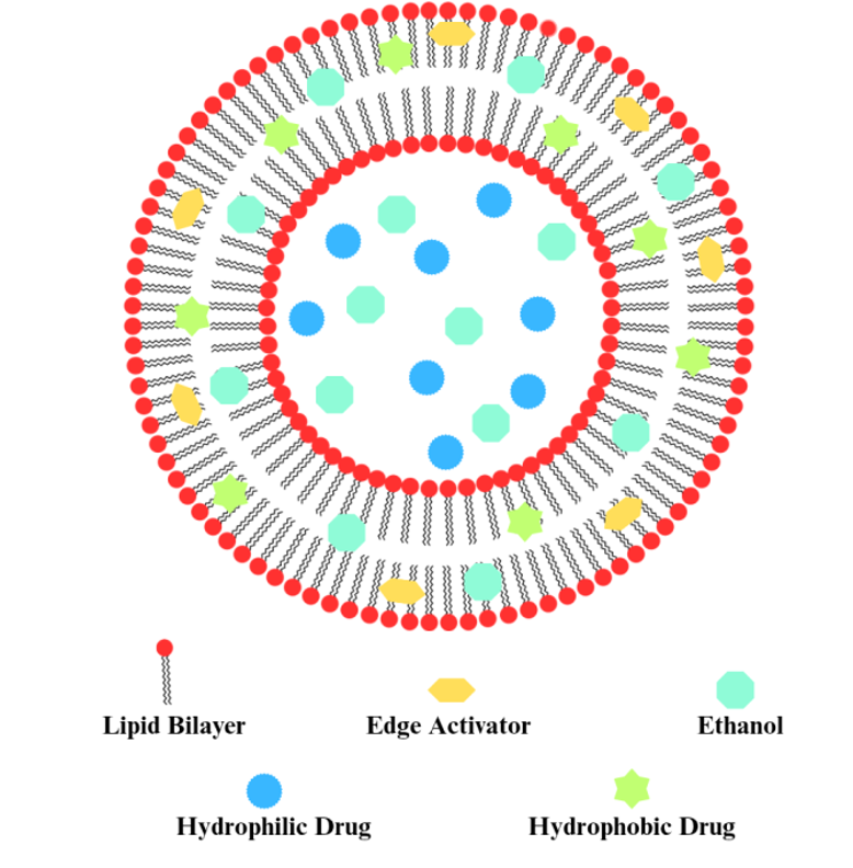
Structurally, transethosomes exhibit an irregular spherical morphology with greater deformability and flexibility than ethosomes and transferosomes, enabling them to regain shape after cellular penetration. Their composition typically includes 2–5% phospholipids, edge activators, 30–40% ethanol, and water, which together facilitate superior skin permeation and efficient drug delivery ⁷². Owing to the biodegradable and biocompatible nature of phospholipids—naturally abundant in eukaryotic membranes—transethosomes provide effective drug encapsulation, avoiding hepatic first-pass metabolism and bypassing the RES system. This makes them suitable for both topical and systemic administration while ensuring improved penetration and safety.
Ethanol and surfactants (edge activators) contribute to membrane softness and enhanced penetration by disrupting stratum corneum lipids, increasing deformability, and facilitating translocation across biological barriers. Cholesterol is often incorporated to improve vesicular stability, regulate drug release kinetics, reduce dosing frequency, and minimize systemic side effects. Moreover, transethosomes can encapsulate both hydrophilic drugs (in the aqueous phase) and hydrophobic drugs (within the phospholipid bilayer).
Surface modifications such as polymeric or carbohydrate coatings can further enhance targeting efficiency. For pulmonary applications, transethosomes improve drug deposition across lung tissues, boosting therapeutic outcomes. Similarly, in the transvaginal route, they circumvent GIT degradation and hepatic first-pass metabolism, thereby enhancing bioavailability and minimizing systemic toxicities. Nonetheless, certain limitations exist, particularly skin irritation due to high ethanol content. Despite this, transethosomes remain scalable and industrially feasible, making them a promising drug delivery option ⁷³,⁷⁴.
Composition of Transethosomes
The composition and architecture of transethosomes are demonstrated below:
a) Phospholipids
Phospholipids, obtained from natural sources (soybean, egg yolk, sunflower) or synthetic ones, are essential for vesicle formation. Natural phospholipids improve skin permeation by fluidizing the stratum corneum, while saturated forms restore barrier function and prolong drug retention. Types include phosphatidic acid (PA), phosphatidylcholine (PC), phosphatidylinositol (PI), phosphatidylethanolamine (PE), phosphatidylserine (PS), and phosphatidylglycerol (PG) ⁷⁵,⁷⁶. Though less stable than synthetic phospholipids such as dimyristoyl phosphatidylcholine (DMPC), distearoyl phosphatidylcholine (DSPC), and dipalmitoyl phosphatidylcholine (DPPC), their concentration (0.5–5%) and type influence size, zeta potential, and entrapment. Ahmed et al. reported multilamellar vesicles (MLVs), while Fang et al. showed unsaturated phospholipids enhanced penetration and entrapment ⁷²,⁷⁷.
b) Ethanol
Ethanol serves a dual function in transethosomes by softening the vesicular membrane for deformability and enhancing skin permeation ⁷⁸. Its concentration (10–50%) influences vesicle size, zeta potential, stability, entrapment efficiency, and penetration. However, higher ethanol levels may excessively solubilize phospholipids, lowering entrapment. Cholesterol addition helps improve flexibility and stability ⁷⁹. Tamer et al. found that 10–20% ethanol yielded better stability than 30–50% ⁸⁰, while Abdulbaqi et al. reported that increasing ethanol from 10% to 30% (w/v) reduced vesicle size, but above this level caused bilayer leakage and size increase ⁸¹.
c) CholesterolIn transethosomal formulations, cholesterol is a key stabilizer that enhances stability and drug entrapment efficiency, though it may also increase vesicle size ⁷⁹. Typically, 3% cholesterol prevents particle agglomeration, maintaining vesicle size, cohesion, and shelf life, making it the most widely used stabilizer. Tamer et al. reported that cholesterol improves vesicle strength and flexibility, especially at high ethanol concentrations, while excess levels reduce encapsulation efficiency due to limited solubility ⁷². Likewise, Cao et al. found that sodium deoxycholate (SDC) above 1% in ivermectin-loaded transethosomes reduced stability and caused drug leakage, lowering therapeutic efficacy ⁸².
d) Edge Activator
Edge activators (EAs) are essential excipients in transethosomal formulations, improving vesicle deformability, flexibility, and permeability. Their type and concentration greatly affect drug delivery, with higher levels enhancing zeta potential, increasing stability, and raising encapsulation efficiency up to threefold due to reduced polarity from deoxycholate–drug ion interactions. Common EAs include Polysorbate 20, sodium cholate, dipotassium glycyrrhizinate, bile salts, Sorbitan laurate 80, 9-Octadecenoic acid, and Polysorbate 80 ⁸³. A study on Fe-chlorophyllin TEs using low EA levels (0.1–0.3% w/v) with phosphatidylcholine and 20% ethanol produced vesicles sized 456–685 nm ⁸⁴.
e) Drug or Active Compound
Incorporation of drugs into transethosomal (TE) systems significantly affects their physicochemical and functional properties, particularly vesicle size, zeta potential, and release behavior, though the polydispersity index remains largely unaffected. Kabil et al. ²⁴ observed that plain nanovesicles carried a negative charge, while drug loading shifted the surface charge to positive. Zhang et al. ⁸⁵ reported that curcumin-loaded TEs showed reduced particle size, higher zeta potential, sustained release up to 48 hours, and superior skin permeation compared to conventional cream. However, Zhuang et al. ⁸⁶ demonstrated that high drug-to-phospholipid ratios reduced stability, promoted drug leakage, and lowered therapeutic efficacy.
f) Surface Functionalization of TEs
Surface modification of transethosomes (TEs) offers a powerful approach to improve targeting, selectivity, and therapeutic efficacy. Functionalization using ligands, peptides, or antibodies enables preferential accumulation at disease sites. For instance, folic acid-functionalized TEs enhanced uptake in folate receptor-rich cancer cells ⁸⁷, while transferrin-conjugated TEs improved delivery in cells with transferrin receptors ⁸⁸,⁸⁹. Peptide-based strategies also showed promise: Ellis and Hicklin ⁹⁰ utilized VEGFR-targeting peptides to enhance anticancer activity, and Zhao et al. ⁹¹ achieved superior uptake in prostate cancer using αvβ3 integrin-targeted TEs. Additionally, antioxidant-coated TEs localized at inflamed tissues, improving drug deformability, encapsulation, and permeation efficiency ⁹¹.The applications and advantages of surface-functionalized TEs are summarized in Figure 4 and Table 4.
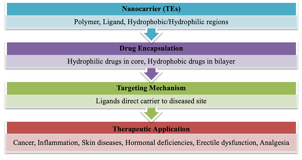
Table 4: Summary of components, roles, and example/application of transethosomes
Component | Role | Example/Application |
Polymer | Stability, long circulation | PEG coating |
Targeting ligand | Cell-specific targeting | Antibodies, peptides |
Hydrophobic drug | Stored in lipid bilayer | Anti-cancer drugs |
Hydrophilic drug | Stored in aqueous core | Hormones, anti-inflammatory drugs |
Application | Wide therapeutic uses | Cancer, Skin diseases, Analgesia |
Mechanism of action of transethosome-mediated skin permeation
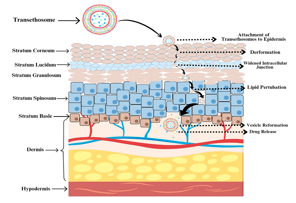
Endogenous molecules cross the stratum corneum (SC) primarily through two routes: the transcellular and intercellular pathways, with the latter serving as the major route for drug permeation into systemic circulation. Drug transport across the skin generally follows Fick’s law of diffusion, indicating a diffusion-driven mechanism ⁹³,⁹⁴. In addition to these, diffusion shunts and appendages such as hair follicles and sweat glands provide alternative pathways for the transport of highly hydrophobic molecules, large biomolecules, and electrolytes. The SC’s lipoidal matrix, supported by its vascular network, is central to maintaining the skin’s barrier properties. However, conventional liposomal formulations face limitations in penetration due to their rigid cholesterol structure, leading to fragmentation and premature drug release in the upper epidermis.
To overcome these barriers, second- and third-generation vesicles have been developed. Transferosomes, with high deformability, migrate from the dry skin surface into deeper hydrated layers, delivering intact vesicles through hydrophilic pores and intercellular channels. Ethosomes, on the other hand, utilize ethanol to impart flexibility and fluidity by interacting with bilayer lipids, thereby enhancing penetration. Transethosomes (TEs) combine the advantages of both systems: they first adhere to the SC, inducing partial dehydration that loosens intracellular junctions, followed by ethanol-mediated deformability that aids deep penetration. Guided by hydration gradients, TEs move from drier to more hydrated layers, ensuring drug transport into systemic circulation. Thus, TE-mediated permeation relies on two mechanisms: (1) ethanol-induced deformability and junction widening and (2) hydration-gradient-driven penetration ⁹⁵,⁹⁶. Figure 5 illustrates the mechanism of action of TE-mediated skin permeation.
Method for preparation and their effect on properties of transethosomes.
Cold Method
The cold method involves preparing transethosomes (TEs) at low temperatures and is widely used due to its simplicity and effectiveness ⁹⁷. In this approach, the aqueous and organic phases are prepared separately. The organic phase is formed by mixing phospholipids, penetration enhancers, and other lipid components in organic solvents at room temperature in a closed vessel ⁹⁸. The aqueous phase such as bidistilled water, standard saline, or buffer solution is then added dropwise at a continuous rate to the organic phase using a syringe pump. The mixture is stirred using a magnetic stirrer for 5–30 minutes at 700–2000 rpm. Studies by Vivek Gupta et al. ⁹⁹ indicated that stirring time directly affects vesicle size. The drug can be dissolved in either the aqueous or organic phase depending on its physicochemical properties ²⁰,²⁸. The final formulation is refrigerated for storage, which helps prevent thermal stress and avoids potential drug degradation ¹⁵,³².
Hot Method
The hot method utilizes heat during TE preparation to enhance lipid solubility, facilitate vesicle formation, and improve drug encapsulation efficiency (EE) ⁹⁷. In this method, the phospholipid colloidal dispersion is prepared in water at 40°C, while simultaneously, a mixture of polyol and ethanol is heated to the same temperature ³⁰. The organic phase is then added to the aqueous phase under continuous stirring to produce a vesicle suspension. Depending on the drug’s solubility, it can be dissolved in water or ethanol. To achieve the desired vesicle size, probe sonication or extrusion can be applied, as demonstrated by Mishra et al. ²⁸.
Ethanol Injection–Sonication Method
The ethanol injection method is employed primarily to produce small unilamellar vesicles with particle sizes in the range of 30–170 nm. Vesicle size is influenced by lipid concentration and injection rate ¹⁰⁰,¹⁰¹. In this technique, phospholipids, surfactants (SA), and the active drug are dissolved in ethanol, which is then injected into an aqueous phase at a controlled flow rate using a syringe. The mixture is subsequently homogenized using an ultrasonic probe ²⁰,¹⁰²⁻¹⁰⁵. Salem et al. ¹⁰⁶ developed an ultrasound-guided injection technique to achieve smaller particle sizes compared to the conventional stirring-guided injection. Singh et al. ¹⁰⁷ combined ethanol injection with the hot method followed by probe sonication to prepare TEs loaded with hydrophilic drugs. They observed that sonicated vesicles exhibited smaller particle sizes than non-sonicated vesicles.
Thin-Film Hydration Technique (TFH)
The thin-film hydration (TFH) technique is a widely used method for preparing transethosomes (TEs). This approach involves forming a thin lipid film on the inner wall of a rotary evaporator flask. The lipid film is then hydrated using water or a buffer solution. To ensure proper bilayer formation, it may be necessary to preheat both the lipid film and the aqueous solution above the lipids’ transition temperature (Tm). Vigorous shaking and, if required, sonication in an ultrasonic bath help detach the film from the flask and form TEs ¹⁰¹,¹⁰⁸.
In this technique, phospholipids, edge activators (EA), and drugs are dissolved in the organic phase within a round-bottom flask. The mixture is then processed in a water bath sonicator until homogeneously distributed. Organic solvents are slowly removed under reduced pressure at a temperature above the lipid Tm using rotary evaporation, forming a thin lipid film on the flask walls ²⁴,¹⁰⁹. To ensure complete removal of residual solvents, the flask is placed in a vacuum oven overnight. The dried lipid film is subsequently hydrated with an aqueous ethanol solution or saline phosphate buffer-ethanol solution while rotating the flask. The resulting TE vesicles are allowed to swell at room temperature and stored at 4–8°C ⁷,²⁴.
Comparative studies: Sonbaty et al. ¹⁰⁵ investigated the impact of TFH and ethanol injection methods on TE quality attributes. The average vesicle size of TEs prepared by TFH was 62.85 nm, compared to 245.3 nm by ethanol injection. The zeta potential and encapsulation efficiency (EE) were 36.3 mV and 82.35% for TFH, versus 49.4 mV and 93.88% for ethanol injection-sonication. Moreover, drug deposition in the skin was 1.5 times higher for TFH-prepared TEs compared to ethanol injection and over 2-fold higher than a drug solution in propylene glycol.
Reverse-Phase Evaporation (REV) Method
The reverse-phase evaporation (REV) method is a less commonly used technique for preparing unilamellar vesicles. This method involves creating a water-in-oil emulsion, followed by evaporation of the organic solvent to form vesicles ¹⁰¹.
In this method, phospholipids, stabilizers, and organic solvents are dried in a round-bottom flask under vacuum to form a thin lipid film. Residual solvents are removed using nitrogen gas. The lipid film is then suspended in a suitable solvent or solvent mixture and stirred at room temperature. The formulation is sonicated for 10 minutes at 5–6°C in a sonication bath to produce a stable emulsion. Removing the solvent under reduced pressure results in colloidal dispersion and formation of the vesicles ²⁸,¹¹⁰,¹¹¹.
Effect on vesicle lamellarity: Nele et al. ¹¹⁰ reported that the preparation method strongly influences the lamellarity of vesicle populations. Specifically:
Film hydration followed by freeze-thaw cycles or agitation on a shaker produces multilamellar, unextruded, or mixed unilamellar/bilamellar vesicles.
The REV method mainly produces unilamellar extruded vesicles, demonstrating its suitability for generating uniform vesicles for specific drug delivery applications.
Characterization of transethosomes
Morphology, Vesicular Size, and Poly-Dispersity Index (PDI)
The most critical parameters in the characterization of transethosomes (TEs) are vesicular size and the poly-dispersity index (PDI). Vesicle size plays a pivotal role in determining the suitability of TEs for inhalation, parenteral administration, and circulation half-life ¹¹². Typically, vesicular carriers are spherical in shape, soft, flexible, and possess an aqueous core. Depending on the formulation, they may be present as small unilamellar or multilamellar vesicles ¹⁵,³². Since most vesicles are in the nanometer range, morphological evaluation is generally performed using transmission electron microscopy (TEM) and scanning electron microscopy (SEM) ¹¹³.
Uniformity in vesicle size is a crucial parameter and can be optimized based on formulation techniques and equipment used. For efficient drug delivery, the preferred vesicle size of TEs generally ranges between 50–200 nm ¹¹⁴. Vesicular size and distribution are commonly determined using photon correlation spectroscopy ⁸⁷, the dynamic light scattering (DLS) method ⁵⁰, and the Microtrac Nanotrac Wave system ⁶.
The PDI value reflects the homogeneity of vesicle populations, describing whether the sample is monodisperse or polydisperse. PDI is dimensionless and ranges between 0 and 1. For drug delivery applications, a PDI of ≤0.3 indicates an appropriate, homogeneous population of vesicles ¹¹⁵. In contrast, higher PDI values suggest heterogeneity and the presence of vesicles with broad size distribution ¹¹⁶. PDI is most commonly measured using DLS, which evaluates the Brownian motion of dispersed particles in solution and their subsequent light scattering ¹¹⁷. The mean particle size is calculated based on the intensity of scattered light ¹¹⁸. Recently, nanoparticle tracking analysis (NTA) has been introduced, which measures vesicular size by tracking the diffusion coefficient of individual vesicles from optical video frames ¹¹⁹. While DLS calculates the diffusion coefficient from intensity fluctuations, NTA determines particle movement directly; therefore, both methods can complement each other in verifying vesicular size measurements.
Zeta Potential
Zeta potential is another critical physicochemical parameter that reflects the stability of TEs. It is usually measured by a Zetasizer ¹¹. The surface charge of vesicular carriers depends largely on the excipients used and significantly influences vesicle stability and interactions. According to Salem et al. ¹, a positive surface charge on vesicles reduces agglomeration due to electrostatic repulsion, thereby enhancing stability. Zeta potential analysis also provides insights into the contribution of individual formulation components, their interactions, and surface chemistry.
In transethosomes, the surface is generally negatively charged due to the presence of ethanol, which enhances colloidal stability ¹²⁰. Additionally, the negative charge may also arise from phosphate groups present in the vesicular structure ¹²¹.
The measurement of zeta potential involves illumination of vesicles with a laser beam, where particle movement under an applied electric field is recorded at a specific angle ¹²². The mobility of TEs is inversely related to their surface charge, and thus zeta potential directly correlates with vesicle stability.
Entrapment Efficiency
A detailed understanding of vesicular properties is essential for optimizing entrapment efficiency (EE) and regulating drug release from transethosomes (TEs). EE is significantly influenced by the synthesis method, composition, and stiffness of the phospholipid bilayer. In the final formulation, both encapsulated and non-encapsulated drug fractions coexist, necessitating the separation of free drug prior to EE determination.
Several methods have been employed for this purpose, including size exclusion chromatography (based on molecular size differences), centrifugation, dialysis membranes with appropriate molecular weight cut-offs, and ultracentrifugation. Among these, ultracentrifugation is widely used to evaluate EE and drug loading. The suspension is centrifuged under controlled temperature and speed to separate the free drug from vesicles without causing structural rupture. The sediment (entrapped drug) and supernatant (free drug) are collected, and vesicles are lysed using a suitable solvent to release the drug. Quantification is generally carried out spectrophotometrically ¹²³.
Entrapment efficiency can be calculated using the following equation:

Drug Content
Drug content represents the quantitative estimation of drug encapsulated within vesicles. For this, vesicles are disrupted to release their contents, which are then analyzed using chromatographic or spectrophotometric methods.
Syed et al. ⁸⁷ reported a validated high-performance liquid chromatographic (HPLC) method employing a ZORBAX Eclipse Plus C18 column with a methanol–phosphate buffer (50 mM) mobile phase to determine benidipine hydrochloride concentration in lipid-based formulations. The method was found to be highly sensitive, accurate, linear, and specific.
Drug content in ethosomal formulations can be expressed as:

Deformability Index
One of the distinctive characteristics of TEs is their deformability, which enables intact passage through skin layers. This property is quantitatively expressed as the deformability index (E):
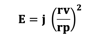
Where:
· E = Deformability index of the vesicle bilayer
· j = Penetration rate through the membrane filter
· rv = Vesicle size after extrusion
· rp = Membrane pore size ³²
Deformability is typically assessed using an extrusion method. It is strongly influenced by the type and concentration of surfactant (SA) incorporated into the formulation ¹⁵.
In Vitro Skin Permeation
In vitro permeation studies are generally performed using a Franz diffusion cell ¹²⁴,¹⁰². A dialysis membrane, often cellophane with a suitable molecular weight cut-off, is selected according to drug characteristics and hydrated with phosphate-buffered saline (PBS, pH 7.4).
The donor compartment is loaded with the formulation.
The receptor compartment is filled with PBS (pH 7.4) and maintained at 32 ± 1 °C with constant stirring (300–400 rpm) to simulate skin conditions.
At predetermined intervals, 1 mL aliquots are withdrawn and replaced with fresh buffer to maintain sink conditions.
Samples are analyzed by UV spectrophotometry ¹⁵,¹⁰².
Stability Studies of Transethosomes (TEs)
The overall performance of TEs depends largely on their chemical and physical stability.
Physical stability is mainly assessed by monitoring vesicle size and appearance. Instability often arises due to aggregation, agglomeration, vesicle fusion, or rupture, which may cause drug leakage during storage.
Chemical stability refers to the ability of TEs to maintain their entrapment efficiency (EE) under varying conditions such as pH fluctuations, changes in electrolyte composition, oxidizing agents, or the presence of surfactants. Any chemical degradation of the lipid bilayer may increase membrane permeability, while drug–phospholipid interactions may further compromise stability.
To ensure stability, formulations are usually stored under controlled conditions. Freshly prepared drug-loaded TEs are kept for 3 months at two different conditions:
Room temperature (25 ± 2 °C, RH 60% ± 5%)
Refrigeration (4 ± 2 °C)
Borosilicate containers are often used to avoid unwanted interactions with the storage vessel. During the study, physical changes and drug content are routinely evaluated to confirm stability ²,⁹⁹,¹²²,¹²⁵.
Therapeutic applications of transethosomes
TEs are recognized for their unique ability to enhance targeted delivery and transport of a wide variety of drugs, regardless of their physicochemical properties. Their ultra-deformable vesicular structure allows them to:
Cross biological barriers efficiently
Provide sustained drug release
Improve drug stability
Minimize systemic side effects through site-specific delivery
Due to these features, TEs are being explored for administration through multiple routes, making them a promising platform for future drug delivery systems. A summary of their therapeutic applications is provided in Table 5.
Table 5: Therapeutic Applications of Transethosomes
S. No | Bioactive Compound / Drug | Method of Preparation | Characteristics (Size / EE% / Zeta Potential / Morphology) | Applications | Ref. |
Anti-fungal drugs | |||||
1 | Voriconazole | Thin-film hydration (TFH) | 191.9 ± 41.5 nm; EE 96.6 ± 2.7%; irregular spherical vesicles | TE significantly enhanced in vivo skin deposition of voriconazole | 20 |
2 | Ketoconazole | TFH technique | 623.06–1532.33 nm; EE 61.45–85.75%; −30 to −40 mV | Showed improved in vivo pharmacodynamic activity compared to conventional liposomes | 18 |
3 | Luliconazole | Ethanol injection with sonication vs. TFH | 246.3 ± 0.56 nm (injection) vs. 62.75 ± 0.16 nm (TFH); EE 82.35 ± 3.68% vs. 93.85 ± 1.78%; ZP 36.3 ± 0.81 vs. 49.4 ± 0.67 mV | TFH method was superior in producing high-quality nanovesicles | 105 |
4 | Econazole nitrate | Homogenization | 159.3 ± 4.3 nm; EE 78.3 ± 2.8%; ZP −27.13 ± 0.33 mV | TE gel showed reduced skin penetration, improved retention, and higher antifungal efficacy compared to marketed cream | 126 |
Anti-inflammatory drugs | |||||
5 | Flurbiprofen | TFH | 152.06–215.2 nm; EE 76.0 ± 0.53%; ZP −30.09 ± 0.46 mV; irregular spheres | Effective transdermal carrier for arthritis management | 12 |
6 | Apremilast | TFH + probe sonication | 89–171 nm; EE 40.32–82.35%; ZP −14.3 mV; spherical vesicles | Improved skin permeation and sustained release | 2 |
7 | Mangiferin | — | 82 & 411 nm; EE 68 ± 3%; spherical and ovoid multilamellar vesicles | Delivered mangiferin effectively to keratinocytes, enhancing antioxidant defense | 127 |
8 | Naproxen sodium | Ethanol injection | 56.94 ± 0.12–291.7 ± 0.09 nm; EE 66.23–93.11%; spherical vesicles | Reduced edema and musculoskeletal pain | 103 |
Anti-hypertensive drugs | |||||
9 | Olmesartan medoxomil | TFH | 222.6 ± 2.5 nm; EE 58.5 ± 1.3%; ZP −20.8 ± 0.3 mV; spherical vesicles | Avoided first-pass metabolism, enhancing transdermal delivery | 128 |
10 | Propranolol HCl | Homogenization | 182.7 ± 5.4 nm; EE 81.98 ± 2.9%; ZP −21.91 ± 0.65 mV; spherical vesicles | Overcame oral drawbacks like first-pass metabolism and toxicity | 129 |
11 | Irbesartan | Cold method | 104.63 ± 0.3–215.06 ± 0.1 nm; EE 76%; ZP −10.7 to −49.3 mV; irregular vesicles | Suitable carrier for poorly soluble drugs | 130 |
12 | Paeonol | Ethanol injection | 122.5 ± 7.5 nm; EE 85.5 ± 5.2%; ZP 13.08 ± 1.18 mV; spherical vesicles | Provided narrow size distribution, prolonged plasma residence, and improved bioavailability | 130 |
Anti-cancer drugs | |||||
13 | Brucine–Strychnine | TFH | 1–100 nm; EE 92.5%; ZP −20 mV; spherical vesicles | Transdermal delivery inhibited HepG2 cell proliferation | 113 |
14 | Rolapitant | TFH | <400 nm; EE 90%; ZP 42.1–51.6 mV; spherical vesicles | Promising tool for lung cancer treatment | 24 |
Anti-aging agents | |||||
15 | Cycloastragenol | — | 100–400 nm; EE 31.5%; spherical vesicles | Potential for anti-aging cosmetic formulations | 7 |
16 | Niacinamide | TFH | 132–215 nm; EE 5.3–7.6%; ZP −21 ± 6 mV; spherical vesicles | TE hydrogels enhanced controlled skin penetration | 5 |
Miscellaneous agents | |||||
17 | Coenzyme Q10 | TFH | 146 ± 26 nm; EE 97.63%; ZP −55 mV; spherical vesicles | Effective topical delivery for androgenic alopecia (AGA) | 4 |
18 | Epigallocatechin-3-gallate | TFH | 153.4–650.8 nm; EE 21.19–58.8%; ZP −2.27 to −5.59 mV | Optimization of TE vesicles is crucial for high entrapment | 130 |
19 | Colchicine | Cold method | 79.8–150.8 nm; EE 60.04–83.57%; ZP ≥ −20 mV; irregular vesicles | Provided alternative to oral route, improving bioavailability | 6 |
20 | Cholecalciferol (Vitamin D3) | Cold method | 182.17 ± 10.17 nm; EE 100%; spherical unilamellar vesicles | Biocompatible and efficient nanocarrier system | 32 |
21 | Tranexamic acid | Cold method | 72 nm; EE 94%; ZP −16 mV | Effective carrier for melasma treatment | 21 |
22 | Progesterone | Injection–sonication | 133.3–349.5 nm; EE 87.93–97.05%; ZP 23.5–74.6 mV; spherical nanovesicles | Increased serum concentration, endometrial thickness, and pregnancy rate | 1 |
23 | 8-Methoxy psoralen | — | 265 ± 2.9 nm; EE 83.87%; nanospheres | Improved narrow-band UVB therapy for vitiligo without side effects | 26 |
24 | Tramadol HCl | Cold method | 149.34–278 nm; EE 79.37%; ZP −22 mV; asymmetrical vesicles | Provided alternative route to bypass low oral bioavailability and adverse effects | 98 |
Conclusion
Transethosomes (TEs) have emerged as an advanced and versatile nanocarrier system that combines the structural benefits of ethosomes and transferosomes, offering superior drug delivery potential through enhanced flexibility, penetration ability, and stability. Their composition—mainly phospholipids, ethanol, surfactants, and penetration enhancers—enables them to encapsulate a wide range of therapeutic agents, both hydrophilic and lipophilic, thereby overcoming the limitations of conventional carriers. Various methods such as cold method, hot method, and ethanol injection with sonication have been optimized for TE preparation, each influencing vesicle size, entrapment efficiency (EE), and deformability. EE, drug loading, and drug content remain critical evaluation parameters, measured through techniques like ultracentrifugation, chromatography, and spectrophotometry, as these directly impact the therapeutic performance of the formulation. Likewise, deformability index and in vitro skin permeation studies using Franz diffusion cells further confirm their ability to traverse skin barriers intact, while stability studies conducted under controlled temperature and humidity highlight challenges such as vesicle fusion, aggregation, drug leakage, and chemical degradation, particularly due to lipid–drug interactions or pH and oxidative changes. Despite these challenges, TEs have demonstrated remarkable potential in improving drug permeation, site-specific targeting, and sustained release, thereby reducing systemic side effects and enhancing patient compliance. Their successful therapeutic applications include delivery of antifungal, anti-inflammatory, cardiovascular, anticancer, and dermatological drugs, along with promising use in cosmetics, nutraceuticals, and hormone therapies, where they enhance bioavailability and treatment efficacy. Taken together, the scientific evidence strongly positions TEs as a novel, adaptable, and clinically valuable drug delivery system capable of addressing multiple challenges in transdermal and targeted delivery. However, translating their laboratory success into real-world applications requires focused research on large-scale production, long-term stability, in vivo performance, clinical trials, and regulatory approval, which will ultimately determine their future as a mainstream strategy in modern pharmaceutics.
References
Salem HF, Kharshoum RM, Abou‐Taleb HA, AbouTaleb HA, AbouElhassan KM. Progesterone-loaded nanosized trans-ethosomes for vaginal permeation enhancement: formulation, statistical optimization, and clinical evaluation in anovulatory polycystic ovary syndrome. J Liposome Res. 2019;29(2):183–194.
Rahangdale M, Pandey P. Development and characterization of apremilast transethosomal gel for transdermal delivery. Int J Pharm Sci Nanotechnol. 2021;14(3):5508–5518.
Nasr M, Wahdan SA. Neuroprotective effects of novel nanosystems simultaneously loaded with vinpocetine and piracetam after intranasal administration. Life Sci. 2019;226:117–129.
El-Zaafarany GM, Abdel-Aziz RTA, Montaser MHA, Nasr M. Coenzyme Q10 phospholipidic vesicular formulations for treatment of androgenic alopecia: ex vivo permeation and clinical appraisal. Expert Opin Drug Deliv. 2021;18(10):1513–1522.
Basto R, Andrade R, Nunes C, Lima SAC, Reis S. Topical delivery of niacinamide to skin using hybrid nanogels enhances photoprotection effect. Pharmaceutics. 2021;13(11):1968.
Abdulbaqi IM, Darwis Y, Abou Assi R, Abdul Karim Khan N. Transethosomal gels as carriers for the transdermal delivery of colchicine: statistical optimization, characterization, and ex vivo evaluation. Drug Des Devel Ther. 2018;12:795–813.
Wang FC, Hudson PL, Burk K, Marangoni AG. Encapsulation of cycloastragenol in phospholipid vesicles enhances transport and delivery across the skin barrier. J Colloid Interface Sci. 2022;608:1222–1228.
Albash R, Abdelbary AA, Refai H, et al. Use of transethosomes for enhancing the transdermal delivery of olmesartan medoxomil: in vitro, ex vivo, and in vivo evaluation. Int J Nanomed. 2019;14:1953–1968.
Verma S, Utreja P. Exploring therapeutic potential of invasomes, transfersomes, transethosomes, oleic acid vesicles, and cubosomes adopting topical/transdermal route. Micro Nanosyst. 2022;14(1):3–20.
Sguizzato M, Ferrara F, Mariani P, et al. Plurethosome as vesicular system for cutaneous administration of mangiferin: formulative study and 3D skin tissue evaluation. Pharmaceutics. 2021;13(8):1124.
Albash R, El-Nabarawi MA, Refai H, et al. Tailoring of PEGylated bilosomes for promoting the transdermal delivery of olmesartan medoxomil: in-vitro characterization, ex-vivo permeation and in-vivo assessment. Int J Nanomed. 2019;14:6555–6574.
Panchaxari Gadad A, Patil AS, Singh Y, Mallappa Dandagi P, Bolmal UB, Basu A. Development and evaluation of flurbiprofen loaded transethosomes to improve transdermal delivery. Indian J Pharm Educ Res. 2020;54(4):954–962.
Guimarães D, Cavaco-Paulo A, Nogueira E. Design of liposomes as drug delivery system for therapeutic applications. Int J Pharm. 2021;601:120571.
Ramkanth S, Anitha P, Gayathri R, Mohan S, Babu D. Formulation and design optimization of nano-transferosomes using pioglitazone and eprosartan mesylate for concomitant therapy against diabetes and hypertension. Eur J Pharm Sci. 2021;162:105811.
Hallan SS, Sguizzato M, Mariani P, et al. Design and characterization of ethosomes for transdermal delivery of caffeic acid. Pharmaceutics. 2020;12(8):740.
Cevc G, Blume G. Lipid vesicles penetrate into intact skin owing to the transdermal osmotic gradients and hydration force. Biochim Biophys Acta Biomembr. 1992;1104(1):226–232.
Hosny KM, Rizg WY, Alhakamy NA, Alamoudi AJ, Mushtaq RY, Safhi AY. Utilization of nanotechnology and experimental design in development and optimization of Aloe vera gel loaded with Finasteride–Garlic Oil–Nanotransfersomes. J Drug Deliv Sci Technol. 2022;68:103130.
Nayak D, Tawale RM, Aranjani JM, Tippavajhala VK. Formulation, optimization and evaluation of novel ultra-deformable vesicular drug delivery system for an anti-fungal drug. AAPS PharmSciTech. 2020;21(5):140.
Nayak D, Thathapudi NC, Ashe S, Nayak B. Bioengineered ethosomes encapsulating AgNPs and tasar silk sericin proteins for non-melanoma skin carcinoma (NMSC) as an alternative therapeutics. Int J Pharm. 2021;596:120265.
Song CK, Balakrishnan P, Shim CK, Chung SJ, Chong S, Kim DD. A novel vesicular carrier, transethosome, for enhanced skin delivery of voriconazole: characterization and in vitro/in vivo evaluation. Colloids Surf B Biointerfaces. 2012;92:299–304.
Shaji J, Parab SS. Formulation development of tranexamic acid loaded transethosomal patch for melasma. Res J Pharm Dosage Forms Technol. 2022;14(1):7–16.
Rodríguez-Luna A, Talero E, Ávila-Román J, et al. Preparation and in vivo evaluation of rosmarinic acid-loaded transethosomes after percutaneous application on a psoriasis animal model. AAPS PharmSciTech. 2021;22(3):103.
Ahmed TA, Alzahrani MM, Sirwi A, Alhakamy NA. Study of the antifungal and ocular permeation of ketoconazole from ophthalmic formulations containing trans-ethosomes nanoparticles. Pharmaceutics. 2021;13(2):151.
Kabil MF, Nasr M, Ibrahim IT, Hassan YA, El-Sherbiny IM. New repurposed rolapitant in nanovesicular systems for lung cancer treatment: development, in vitro assessment and in vivo biodistribution study. Eur J Pharm Sci. 2022;171:106119.
Esentürk güzel I. Improved skin penetration and deposition of naftifine from transethosomes and transethosomal gel formulations. Farmacia. 2022;70(3):514–521.
Mahmoud DB, ElMeshad AN, Fadel M, Tawfik A, Ramez SA. Photodynamic therapy fortified with topical oleyl alcohol-based transethosomal 8-methoxypsoralen for ameliorating vitiligo: optimization and clinical study. Int J Pharm. 2022;614:121459.
Ansari MD, Ahmed S, Imam SS, et al. CCD based development and characterization of nano-transethosome to augment the antidepressant effect of agomelatine on Swiss albino mice. J Drug Deliv Sci Technol. 2019;54:101234.
Mishra KK, Kaur CD, Gupta A. Development of itraconazole loaded ultra-deformable transethosomes containing oleic acid for effective treatment of dermatophytosis: Box-Behnken design, ex vivo and in vivo studies. J Drug Deliv Sci Technol. 2022;67:102998.
Al-Daraji M, Samy N, Fadel M. Topical indocyanine green nano system photodynamic therapy versus cryotherapy for treatment of common warts: a randomized controlled study. J Nat Remedies. 2021;22(1–2):157–164.
Youssef A, Dudhipala N, Majumdar S. Ciprofloxacin loaded nanostructured lipid carriers incorporated into in situ gels to improve management of bacterial endophthalmitis. Pharmaceutics. 2020;12(6):572.
Hesham H, Rady M, Hathout RM, Abdel-Halim M, Mansour S. The skin delivery of tofacitinib citrate using transethosomes and hybridized ethosomes/nanostructured lipid carriers for vitiligo therapy: dermatopharmacokinetics and in vivo assays. Int J Pharm. 2022;629:122387
Costanzo M, Esposito E, Sguizzato M, et al. Formulative study and intracellular fate evaluation of ethosomes and transethosomes for vitamin D3 delivery. Int J Mol Sci. 2021;22(10):5341.
Ansari SA, Qadir A, Warsi MH, et al. Ethosomes-based gel formulation of karanjin for treatment of acne vulgaris: in vitro investigations and preclinical assessment. 3 Biotech. 2021;11(11):456.
Ahmed TA. Study of the pharmacokinetics, pharmacodynamics and hepatoprotective activity of rosuvastatin from drug-loaded lyophilized orodispersible tablets containing transfersomes nanoparticles. J Drug Deliv Sci Technol. 2021;63:102489.
Mosallam S, Albash R, Abdelbari MA. Advanced vesicular systems for antifungal drug delivery. AAPS PharmSciTech. 2022;23(6):206.
Honeywell-Naguyen PL, Bouwstre JA. Vesicles as a tool for transdermal delivery. Drug Discov Today Technol. 2005;2(1):67–74.
Dhopavkar S, Karu P. Transferosomes – a boon for transdermal delivery. Indo Am J Pharm Sci. 2017;4(9):2908–2919.
Shaji J, Bajaj R. Transethosomes: a new prospect for enhanced transdermal delivery. Int J Pharm Sci Res. 2018;9(7):2681–2685.
Bajaj K, Parab B, Shidhaye S. Nano-transethosomes: a novel tool for drug delivery through skin. Indian J Pharm Educ Res. 2021;1–10.
Shaji J, Bajaj R. Formulation development of 5-Fluorouracil transethosomes for skin cancer therapy. Int J Pharm Pharm Res. 2017;11(1):454–464
Tiwari A, Mishra MK, Nayak K, Yadav SK, Shukla A. Ethosomes: a novel vesicular carrier system for therapeutic applications. IOSR J Pharm. 2016;6(9):25–33.
Sundar VD, Divya P, Dhanaraju MD. Design, development and characterisation of tramadol hydrochloride loaded transethosomal gel formulation for effective pain management. Indian J Pharm Educ Res. 2020;54(2s):s88–97.
Jayaprakash R, Hameed J, Anupriya A. An overview of transdermal delivery system. Asian J Pharm Clin Res. 2017;10(10):36–40.
Gondkar SB, Patil NR, Saudagar RB. Formulation development and characterization of drug loaded transethosomes for transdermal delivery: review article. Int J ChemTech Res. 2017;10(6):535–44.
Chaurasiya P, Ganju E, Upmanyu N, Ray SK, Jain P. Transferosomes: a novel technique for transdermal drug delivery. J Drug Deliv Ther. 2019;9(1):279–85.
Tanner T, Marks R. Delivering drugs by the transdermal route: review and comment. Skin Res Technol. 2008;14:249–60.
Vaseem RS, D'Cruz A, Shetty S, H V, Vardhan A, R SS, et al. Transdermal drug delivery systems: a focused review of the physical methods of permeation enhancement. Adv Pharm Bull. 2023.
Yu YQ, Yang X, Wu XF, Fan YB. Enhancing permeation of drug molecules across the skin via delivery in nanocarriers: novel strategies for effective transdermal applications. Front Bioeng Biotechnol. 2021;9.
Ramadon D, McCrudden MTC, Courtenay AJ, Donnelly RF. Enhancement strategies for transdermal drug delivery systems: current trends and applications. Drug Deliv Transl Res. 2022;12:758–91.
Jäger J, Vahav I, Thon M, Waaijman T, Spanhaak B, de Kok M, et al. Reconstructed human skin with hypodermis shows essential role of adipose tissue in skin metabolism. Tissue Eng Regen Med. 2024;21:499–511.
Phatale V, Vaiphei KK, Jha S, Patil D, Agrawal M, Alexander A. Overcoming skin barriers through advanced transdermal drug delivery approaches. J Control Release. 2022;351:361–80.
Marwah H, Garg T, Goyal AK, Rath G. Permeation enhancer strategies in transdermal drug delivery. Drug Deliv. 2016;23:564–78.
Majumdar S, Mahanti B, Kar AK, Parya H, Ghosh A, Kar B. Nanoliposome: as a smart nanocarrier in transdermal drug delivery system. Intell Pharm. 2024.
Kumar R, Dkhar DS, Kumari R, Divya, Mahapatra S, Dubey VK, et al. Lipid based nanocarriers: production techniques, concepts, and commercialization aspect. J Drug Deliv Sci Technol. 2022;74:103526.
Lu H, Zhang S, Wang J, Chen Q. A review on polymer and lipid-based nanocarriers and its application to nano-pharmaceutical and food-based systems. Front Nutr. 2021;8.
Souto EB, Macedo AS, Dias-Ferreira J, Cano A, Zielińska A, Matos CM. Elastic and ultradeformable liposomes for transdermal delivery of active pharmaceutical ingredients (APIs). Int J Mol Sci. 2021;22:9743.
Richard C, Cassel S, Blanzat M. Vesicular systems for dermal and transdermal drug delivery. RSC Adv. 2021;11:442–51
Nayak D, Tippavajhala VK. A comprehensive review on preparation, evaluation and applications of deformable liposomes. Iran J Pharm Res. 2021;20:186–205.
Tiwari G, Tiwari R, Singh R, Rai AK. Ultra-deformable liposomes as flexible nanovesicular carrier to penetrate versatile drugs transdermally. Nanosci Nanotechnol Asia. 2020;10:12–20.
Zeb A, Arif ST, Malik M, Shah FA, Din FU, Qureshi OS, et al. Potential of nanoparticulate carriers for improved drug delivery via skin. J Pharm Investig. 2019;49:485–517.
Andra VVSNL, Pammi SVN, Bhatraju LVKP, Ruddaraju LK. A comprehensive review on novel liposomal methodologies, commercial formulations, clinical trials and patents. Bionanoscience. 2022;12:274–91.
Bhardwaj P, Tripathi P, Gupta R, Pandey S. Niosomes: a review on niosomal research in the last decade. J Drug Deliv Sci Technol. 2020;56:101581.
Emanet M, Ciofani G. Ethosomes as promising transdermal delivery systems of natural-derived active compounds. Adv NanoBiomed Res. 2023;3:e2200151.
Fernández-García R, Lalatsa A, Statts L, Bolás-Fernández F, Ballesteros MP, Serrano DR. Transferosomes as nanocarriers for drugs across the skin: quality by design from lab to industrial scale. Int J Pharm. 2020;573:118817.
Munir M, Zaman M, Waqar MA, Hameed H, Riaz T. A comprehensive review on transethosomes as a novel vesicular approach for drug delivery through transdermal route. J Liposome Res. 2024;34(2):203–18.
Wang L, Huang X, Jing H, Ma C, Wang H. Bilosomes as effective delivery systems to improve the gastrointestinal stability and bioavailability of epigallocatechin gallate (EGCG). Food Res Int. 2021;149:110631.
Kumar B, Pandey M, Aggarwal R, Sahoo PK. A comprehensive review on invasomal carriers incorporating natural terpenes for augmented transdermal delivery. Future J Pharm Sci. 2022;8:50.
Arundhasree, Rajalakshmi R, Aiswarya R, Abhirami, Rajendra Kumar, Sreelakshmi S, Kumar S, et al. Ufasomes: unsaturated fatty acid-based vesicular drug delivery system. Int J Appl Pharm. 2021;13(1):76–83.
Barani M, Sangiovanni E, Angarano M, Rajizadeh MA, Mehrabani M, Piazza S, et al. Phytosomes as innovative delivery systems for phytochemicals: a comprehensive review of literature. Int J Nanomedicine. 2021;16:6983–7022.
El-Zaafarany GM, Abdel-Aziz RTA, Montaser MHA, Nasr M. Coenzyme Q10 phospholipidic vesicular formulations for treatment of androgenic alopecia: ex vivo permeation and clinical appraisal. Expert Opin Drug Deliv. 2021;18(11):1513–22.
Nasr M, Wahdan SA. Neuroprotective effects of novel nanosystems simultaneously loaded with vinpocetine and piracetam after intranasal administration. Life Sci. 2019;226:117–29.
Chowdary P, Padmakumar A, Rengan AK. Exploring the potential of transethosomes in therapeutic delivery: a comprehensive review. MedComm Biomater Appl. 2023;2:e38.
Raj A, Dua K, Nair RS, Chandran CS, Alex AT. Transethosome: an ultra-deformable ethanolic vesicle for enhanced transdermal drug delivery. Chem Phys Lipids. 2023;255:105315.
Adnan M, Haider MF, Naseem N, Haider T. Transethosomes: a promising challenge for topical delivery. Drug Res. 2023;73(5):200–12.
Li J, Wang X, Zhang T, Wang C, Huang Z, Luo X, et al. A review on phospholipids and their main applications in drug delivery systems. Asian J Pharm Sci. 2015;10(2):81–98.
Sakdiset P, Okada A, Todo H, Sugibayashi K. Selection of phospholipids to design liposome preparations with high skin penetration-enhancing effects. J Drug Deliv Sci Technol. 2018;44:58–64.
Ahmed TA. Study the pharmacokinetics, pharmacodynamics and hepatoprotective activity of rosuvastatin from drug loaded lyophilized orodispersible tablets containing transfersomes nanoparticles. J Drug Deliv Sci Technol. 2021;63:102489.
Gupta R, Badhe Y, Rai B, Mitragotri S. Molecular mechanism of the skin permeation enhancing effect of ethanol: a molecular dynamics study. RSC Adv. 2020;10:12234–48.
Nsairat H, Ibrahim AA, Jaber AM, Abdelghany S, Atwan R, Shalan N, et al. Liposome bilayer stability: emphasis on cholesterol and its alternatives. J Liposome Res. 2024;34(2):178–202.
Tamer TM. Formulation and optimization of a new transethosomal gel containing meloxicam: in vitro, in vivo evaluation and gastrointestinal toxicity studies. Int J Pharm. 2020;583:119402.
Abdallah HH. Development and characterization of transethosomes for enhanced skin delivery of 5-fluorouracil. Drug Dev Ind Pharm. 2019;45(4):620–9.
Cao Y. Transethosomes containing sodium deoxycholate for dermal delivery of ivermectin: optimization, characterization, and in vitro/in vivo evaluation. Drug Des Devel Ther. 2019;13:2465–78.
Natsheh H, Touitou E. Phospholipid vesicles for dermal/transdermal and nasal administration of active molecules: the effect of surfactants and alcohols on the fluidity of their lipid bilayers and penetration enhancement properties. Molecules. 2020;25(13):2959.
Rady M, Gomaa I, Afifi N, Abdel-Kader M. Dermal delivery of Fe-chlorophyllin via ultradeformable nanovesicles for photodynamic therapy in melanoma animal model. Int J Pharm. 2018;548(1):480–90.
Zhuang Y. Effects of drug to phospholipid ratios on the physicochemical properties of transethosomes as dermal carriers. J Nanosci Nanotechnol. 2014;14(2):1359–65.
Torchilin VP. Recent advances with liposomes as pharmaceutical carriers. Nat Rev Drug Discov. 2005;4(2):145–60.
Limongi T, Susa F, Marini M, et al. Lipid-based nanovesicular drug delivery systems. Nanomaterials. 2021;11(12):3391.
Mishra DK, Shandilya R, Mishra PK. Lipid based nanocarriers: a translational perspective. Nanomedicine. 2018;14(7):2023–50.
Ellis LM, Hicklin DJ. VEGF-targeted therapy: mechanisms of anti-tumour activity. Nat Rev Cancer. 2008;8(8):579–91.
Zhao M, Ding J, Mao Q, et al. A novel αvβ3 integrin-targeted NIR-II nanoprobe for multimodal imaging-guided photothermal therapy of tumors in vivo. Nanoscale. 2020;12(13):6953–8.
Song H, Wen J, Li H, et al. Enhanced transdermal permeability and drug deposition of rheumatoid arthritis via sinomenine hydrochloride-loaded antioxidant surface transethosome. Int J Nanomedicine. 2019;14:3177–88.
Hansen S, Lehr CM, Schaefer UF. Improved input parameters for diffusion models of skin absorption. Adv Drug Deliv Rev. 2013;65(2):251–64.
Wu L, Shrestha P, Iapichino M, Cai Y, Kim B, Stoeber B. Characterization method for calculating diffusion coefficient of drug from polylactic acid (PLA) microneedles into the skin. J Drug Deliv Sci Technol. 2021;61:102192.
Bajaj KJ, Parab BS, Shidhaye SS. Nano-transethosomes: a novel tool for drug delivery through skin. Indian J Pharm Educ Res. 2021;55:s1–10.
Joga R, Kannan B, Yerram S, Nandagawale A, Gawai M, Behera HK, et al. Transethosome as a versatile nano vehicle for various indications and its regulatory insights. Pharm Dev Technol. 2023;28(9):1056–78.
Shah S, Dhawan V, Holm R, Nagarsenker MS, Perrie Y. Liposomes: advancements and innovation in the manufacturing process. Adv Drug Deliv Rev. 2020;154–155:102–22.
Sundar VD, Divya P, Dhanaraju MD. Design, development and characterisation of tramadol hydrochloride loaded transethosomal gel formulation for effective pain management. Indian J Pharm Educ Res. 2020;54(2s):s88–97.
Gupta V, Joshi NK. Formulation, development and evaluation of ketoprofen loaded transethosomes gel. J Drug Deliv Ther. 2022;12(1):86–90.
Abdulbaqi IM, Darwis Y, Khan NK, et al. Ethosomal nanocarriers: the impact of constituents and formulation techniques on ethosomal properties, in vivo studies, and clinical trials. Int J Nanomedicine. 2016;11:2279–304.
Šturm L, Poklar Ulrih N. Basic methods for preparation of liposomes and studying their interactions with different compounds, with the emphasis on polyphenols. Int J Mol Sci. 2021;22(12):6547.
Pérez J, Cortés DM, Gómez Y. Potential use of transethosomes as a transdermal delivery system for metabolites from Chenopodium murale. Mater Today Commun. 2022;30:103165.
Kaul S, Jain N, Nagaich U. Ultra deformable vesicles for boosting transdermal delivery of 2-arylpropionic acid class drug for management of musculoskeletal pain. J Pharm Investig. 2022;52(2):217–31.
El-Sonbaty MM, Akl MA, El-Say KM, Kassem AA. Does the technical methodology influence the quality attributes and the potential of skin permeation of luliconazole loaded transethosomes? J Drug Deliv Sci Technol. 2022;68:103096.
Salem HF, Nafady MM, Kharshoum RM, et al. Mitigation of rheumatic arthritis in a rat model via transdermal delivery of dapoxetine HCl amalgamated as a nanoplatform: in vitro and in vivo assessment. Int J Nanomedicine. 2020;15:1517–35.
Singh M, Lee KE, Vinayagam R, Kang SG. Synthesis and assessment of lipid nanovesicles for efficient transdermal delivery of hydrophilic molecules. Nano. 2022;17(4):2250031.
Fu Y, Saraswat A, Vartak R, et al. Liposomal formulation. In: Multifunctional Nanocarriers. Elsevier; 2022. p.79–102.
Khan AU, Jamshaid H, Din F, et al. Designing, optimization and characterization of trifluralin transfersomal gel to passively target cutaneous leishmaniasis. J Pharm Sci. 2022;111(16):1798–811.
Nele V, Holme MN, Kauscher U, Thomas MR, Doutch JJ, Stevens MM. Effect of formulation method, lipid composition, and pegylation on vesicle lamellarity: a small-angle neutron scattering study. Langmuir. 2019;35(18):6064–74.
Zare Kazemabadi F, Heydarinasab A, Akbarzadeh A, Ardjmand M. Preparation, characterization and in vitro evaluation of PEGylated nanoliposomal containing etoposide on lung cancer. Artif Cells Nanomed Biotechnol. 2019;47(1):3222–30.
Elsana H, Olusanya TOB, Carr-Wilkinson J, Darby S, Faheem A, Elkordy AA. Evaluation of novel cationic gene-based liposomes with cyclodextrin prepared by thin film hydration and microfluidic systems. Sci Rep. 2019;9(1):15120.
Wang Y, Wang R, Qi X, et al. Novel transethosomes for the delivery of brucine and strychnine: formulation optimization, characterization and in vitro evaluation in hepatoma cells. J Drug Deliv Sci Technol. 2021;64:102425.
William B, Noémie P, Brigitte E, Géraldine P. Supercritical fluid methods: an alternative to conventional methods to prepare liposomes. Chem Eng J. 2020;383:123106.
Danaei M, Dehghankhold M, Ataei S, et al. Impact of particle size and polydispersity index on the clinical applications of lipidic nanocarrier systems. Pharmaceutics. 2018;10(2):57.
Gaumet M, Vargas A, Gurny R, Delie F. Nanoparticles for drug delivery: the need for precision in reporting particle size parameters. Eur J Pharm Biopharm. 2008;69(1):1–9.
Şahin Bektay H, Kahraman E, Güngör S. Supplement 1. Maced Pharm Bull. 2020;66(3):101–2.
Isalomboto Nkanga C, Murhimalika Bapolisi A, Ikemefuna Okafor N, et al. General perception of liposomes: formation, manufacturing and applications. In: Liposomes – Advances and Perspectives. IntechOpen; 2019.
Kim A, Ng WB, Bernt W, Cho NJ. Validation of size estimation of nanoparticle tracking analysis on polydisperse macromolecule assembly. Sci Rep. 2019;9(1):2639.
Mbah CC, Builders PF, Attama AA. Nanovesicular carriers as alternative drug delivery systems: ethosomes in focus. Expert Opin Drug Deliv. 2014;11(1):45–59.
Abdellatif AAH, Aldosari BN, Al-Subaiyel A, et al. Trans-ethosomal gel for the topical delivery of celecoxib: formulation and estimation of skin cancer progression. Pharmaceutics. 2022;15(1):22.
Laouini A, Jaafar-Maalej C, Limayem-Blouza I, Sfar S, Charcosset C, Fessi H. Preparation, characterization and applications of liposomes: state of the art. J Colloid Sci Biotechnol. 2012;1(2):147–68.
Bi Y, Xia H, Li L, et al. Liposomal vitamin D3 as an anti-aging agent for the skin. Pharmaceutics. 2019;11(7):311.
Sudhakar K, Mishra V, Jain S, Rompicherla NC, Malviya N, Tambuwala MM. Development and evaluation of the effect of ethanol and surfactant in vesicular carriers on lamivudine permeation through the skin. Int J Pharm. 2021;610:121226.
Maherani B, Arab-Tehrany E, Mozafari MR, Gaiani C, Linder M. Liposomes: a review of manufacturing techniques and targeting strategies. Curr Nanosci. 2011;7(3):436–52.
Verma S, Utreja P. Transethosomes of econazole nitrate for transdermal delivery: development, in-vitro characterization, and ex-vivo assessment. Pharm Nanotechnol. 2018;6(3):171–9.
Sguizzato M, Ferrara F, Hallan SS, et al. Ethosomes and transethosomes for mangiferin transdermal delivery. Antioxidants (Basel). 2021;10(5):768.
Albash R, Abdelbary AA, Refai H, et al. Use of transethosomes for enhancing the transdermal delivery of olmesartan medoxomil: in vitro, ex vivo, and in vivo evaluation. Int J Nanomedicine. 2019;14:1953–68.
Kumar L, Utreja P. Formulation and characterization of transethosomes for enhanced transdermal delivery of propranolol hydrochloride. Micro Nanosyst. 2020;12(1):38–47.
Verma A, Mishra M. Formulation and characterization of transethosomal-loaded nanoparticles of irbesartan. Int J Pharm Res Scholars. 2021;10(1):28–35.
Chen ZX, Li B, Liu T, et al. Evaluation of paeonol-loaded transethosomes as transdermal delivery carriers. Eur J Pharm Sci. 2017;99:240–5.
El Kayal M. Optimization of the colloidal properties of different vesicular systems aiming to encapsulate (-)-epigallocatechin-3-gallate. Farmacia. 2020;68(1):97–110.
Authors
Corresponding Author: Dr. Akanksha Dwivedi
School of Pharmacy and Technology Management, SVKM's, NMIMS,
Indore-453112, Madhya Pradesh, India.
Email: Akanksha.Dwivedi@nmims.edu
Telephone: +91 99938-40688
Daniya Khan, Akanksha Dwivedi*, G. N. Darwhekar
School of Pharmacy and Technology Management





Comments