- lauraclark849
- 2 days ago
- 21 min read
Peer Review Article | Open Access | Published 2 July 2026
Expanding test panels for pharmaceutical microbiology: Making an informed decision
Tim Sandle, Ph.D., CBiol, FIScT | EJPPS | 3102 (2026) | https://doi.org/10.37521/ejpps31203
Abstract
Expanding microbial test panels beyond pharmacopeial strains is increasingly important for ensuring the robustness and relevance of pharmaceutical microbiology methods. This paper presents a structured, data-driven approach to selecting representative organisms, drawing on environmental monitoring data from cleanrooms, aseptic processing, water systems, and in-process samples. While the use of environmental isolates can improve representativeness, their variability and lack of standardisation require careful consideration. The study demonstrates that selection should be based on frequency of recovery, morphological grouping, ecological relevance, and phylogenetic diversity, rather than simply origin. By comparing facility isolates with compendial organisms, the methodology ensures supplementary strains provide meaningful challenge without redundancy. The outcome is a rationalised panel of additional organisms that better reflects facility microbiota and supports applications such as growth promotion testing, disinfectant efficacy studies, and method validation. This structured approach enables scientifically justified, reproducible, and risk-based decisions when expanding microbiological test panels.
Introduction
In 2022, this author wrote two articles about environmental isolates for EJPPS. The first of these – “Walk on the wild side: The application of environmental isolates in microbiological testing” questioned whether the use of environmental isolates to challenge microbial test methods and to release culture media adds value. This review concluded that there is considerable merit in broadening the test panel beyond pharmacopeial recommended strains; however, there is little in favour of using actual organisms isolated from the manufacturing area. This is because the ‘wildtype’ characteristics were unlikely to be retained at a level of any phenotypic value (and, conversely, if these properties were retained, this would probably make them unreliable as control strains) ¹.
This was then confirmed by the follow-up paper, “Examination of the growth rates of environmental isolates compared with compendial strains”. This paper presented experimental data where the growth rates of compendial strains sourced from a culture collection were compared with manufacturing facility isolates. The research found that environmental isolates do take slightly longer to grow compared with laboratory strains, yet this time difference was within the recommended incubation times of each test type. Since the physiological state of an organism will influence the triggers necessary to move out of a state of dormancy, as well as the speed of the awakening processes once the organism encounters the nutrients and environmental conditions necessary for growth ², potentially some metabolically influenced effect was observed ³. Therefore, microbiologists should generally expect slower growth, but test methods do not need to be adapted to compensate. Hence, there is no strong argument in favour of using facility strains (although equally, there is nothing explicitly wrong with doing so).
This paper forms an approximate trilogy, returning to the subject of environmental isolates but with a focus on how to select representative organisms, be they actual isolates or organisms sourced from culture collections to broaden the laboratory test panel.
Expanding the test panel of microorganisms to assess test reliability is important. Yet the importance is only logical if the organisms are sufficiently, indeed genetically, different.
Microbial test panels
The test panels of organisms recommended by compendia and standards are in place to allow for reproducibility between laboratories. In the case of most standards, these are designed to be multi-industry. Thus, the range of isolates quoted may or may not be suitable for the application. One role of the microbiologist is to review the panel and decide whether to supplement it with additional isolates. There are several applications where this might be warranted, including:
Disinfectant challenge tests (disinfectant efficacy tests).
Growth promotion tests for new lots of culture media.
Antimicrobial effectiveness test (or preservative efficacy test)
Method validation.
The reasons for including additional organisms in microbiological tests can be divided between those that involve expanding the test panel of organisms with additional organisms and those that relate to comments made by regulators. The primary reasons for including environmental isolates are:
To widen the test panel, to make the challenge organisms used more representative of what is being recovered from the environment or samples ⁴.
To include types of organisms that are not fully represented in the test panel ⁵.
To add organisms that match regulatory concerns (a representative of the Burkholderia cepacia complex is an example of a concern highlighted by the U.S. FDA in 2017).
The question is then, which the first paper sought to address, whether these additional organisms needed to have been isolated from the actual facility and preserved in the laboratory as test isolates or purchased from a recognised culture collection.
A regulatory expectation that environmental isolates are incorporated into microbiology testing gained momentum during the late 1990s, and this has largely become commonplace “Discussions at conferences and symposia suggest that US and European regulators are asking whether environmental isolates are included in release tests and validation and are expecting a justification if this is not the case.” ⁶ This is supported by Booth, who reviewed several FDA warning letters where citations were issued for firms failing to use environmental isolates for media release testing ⁷. Yet environmental isolates cannot be standardised, and therefore the test methods developed and used across different laboratories will not be comparable since different organisms have been used ⁸.
The argument about the origin of the organisms, as a dialogue between agencies, is one for another time. This paper proceeds to the topic at hand – organism selection.
Methodology
To seek appropriate environmental isolates, different parts of the pharmaceutical facility will have different types of organisms. What is found in a purified water system will be, for example, different to what is found in a cleanroom. Therefore, the data sets need to be appropriately divided. Within a system, there will be different types of organisms occurring within morphological groups, with morphology determined by staining for bacteria and between yeasts and moulds (filamentous) for fungi. Bacterial morphology refers to the size, shape, structure, and arrangement of bacterial cells. It is one of the most fundamental characteristics used to identify and classify bacteria. To this, cell wall staining can be added. This produces:
Gram-positive cocci.
Gram-positive rods (which can be further subdivided into spore formers and non-spore formers).
Gram-negative rods.
Gram-negative cocci (if any were to occur in significant numbers).
Other forms, such as spiral forms (helical or curved), Vibrio (comma-shaped), filamentous (thread-like formations), and pleomorphic (organisms that present variable shapes), are unlikely to be detected in sufficiently high numbers.
The reason why this is a useful point for categorisation is that microbial morphologies are an effective way to divide organisms from different potential origins (human skin, environment, water, etc.), and they reflect the diversity of microbial types that tend to occur across contamination investigations. Twinned together, these reasons ensure representativeness through understanding typical microbial populations and ecology.
Furthermore, adopting a consistent approach and conducting annual screening enables the microbiologist to assess:
New/unexpected colony types,
Changes in growth patterns over time,
Potential emerging contamination issues,
Understanding the ‘normal’ microbiota.
Therefore, morphology is useful for grouping environmental isolates using a simple method of categorisation, but one which also ensures representative coverage of facility microbiota.
Data for each assessed area was collected for the period 1st January 2025 to 31st January 2025 into different systems:
Group 1: Grade C and D cleanrooms (described as ‘process areas’)
Group 2: Aseptic processing
Group 3: Water systems
Group 4: Intermedia product bioburden
Group 1: Process areas
The data for process areas (Grade C and D cleanrooms) showed the following pattern, presented by morphological type (Figure 1):

The organisms were then divided by genus, which produced:

Across this dataset, the genera and species that appear most frequently were:
Kocuria (salsicia, rhizophila, palustris, carniphila/atrinae)
Staphylococcus (equorum, epidermidis, hominis, capitis, warneri, aureus)
Bacillus (cereus group, licheniformis, pumilus, altitudinis, safensis)
Serratia (grimesii, liquefaciens, proteamaculans)
Psychrobacter (faecalis, pulmonis, alimentarius)
Rhodococcus (erythropolis)
Micrococcus (luteus)
Paenibacillus (glucanolyticus/lautus)
In terms of the most common organisms by morphology, the output is presented in Table 1:
Table 1: Most common process area organisms by morphology
Morphology | Proportion of isolates | Most common genera | Most common species (representative of the most common genera) |
Gram-positive coccus | ~40% | Kocuria | K. rhizophilia |
Gram-positive rod (non-sporing) | ~17% | Microbacterium | No specific species dominates |
Gram-positive rod (sporing) | ~11% | Bacillus | B. cereus |
Gram-negative rod | ~25% | Pseudomonas | Ps. fluorescens or Ps. stutzeri |
Fungus | ~5% | Aspergillus | A.fumigatus |
In terms of the selection, Kocuria was the largest genus of Gram-positive cocci (at 17% of isolates), followed by Staphylococci at 11%, and Micrococcus at 5%. Therefore, the most common species of Kocuria was selected. K. rhizophilia represented 25% of the species within this genus. Kocuria live on human skin and in the oral cavity ⁹, as well as being found in soil and the general environment; there are close similarities with Micrococcus (being members of the family Micrococcaceae). K. rhizophilia is a soil-dwelling Gram-positive bacterium ¹⁰; it also inhabits normal human skin, particularly in areas with higher sebum, and is generally considered part of the healthy skin microbiome. Its presence in cleanrooms, to a degree, is not unexpected.
With Gram-positive non-sporing rods, Microbacterium was the largest genus, accounting for ~40% of this morphological group. Of these organisms, M. liquefaciens accounted for 26% of the isolates. Microbacteria are associated with many raw materials, as well as the surrounding environment ¹¹. They can be short-term residents on the human body. M. liquefaciens falls into both areas, in terms of potential origins ¹².
Of the spore-forming Gram-positive rods, Bacillus represented the largest genus, and the most commonly isolated species is B. cereus. This organism is commonly found in soil, and its presence in cleanrooms can be problematic, especially when it enters aseptic processing areas ¹³ due to its relative resistance to more extreme conditions (temperature, lack of nutrients, UV light, etc.). The bacterium is also an early coloniser of biofilm communities ¹⁴.
With Gram-negative rods, the most common genera are Pseudomonas (40% of Gram-negative rods), followed by Chryseobacterium at 15%. Of the Pseudomonas species, the predominant organism is Ps. fluorescens ¹⁵. This organism is capable of accelerated growth in the cold, making it unsuitable for pre-preparing cultures for dilution. The second most common organism is Ps. stutzeri.
Group 2: Aseptic Filling Suite
For the second area, this relates to aseptic processing areas (Grade A and B areas, albeit with almost all isolates coming from Grade B). For 2025, there were 716 identifications performed.
In terms of the most common organisms by morphology, these are displayed in Table 2:
Table 2: Most common aseptic area organisms by morphology
Morphology | Proportion of isolates | Most common genera | Most common species (representative of the moist common genera) |
Gram-positive coccus* | ~73% | Micrococcus | M. luteus |
Gram-positive rod (non-sporing) | ~11% | Brevibacterium | B. pityocampae or C. tuberculostearicum |
Gram-positive rod (sporing) | ~2% | Bacillus | B. subtilis |
Gram-negative rod | ~10% | Moraxella | M. osleonsis |
Fungus | ~3% | Penicillium | P. chrysogenum |
To derive the above outcomes, the following data set was used:
Gram-positive cocci
The division between the major Gram-positive cocci genera was:
210 = Micrococcus
198 = Staphylococcus
97 = Kocuria
Of these organisms, the most common species was Micrococcus luteus at 198 isolates. M. luteus is abundant on human skin ¹⁶. The organism is also known for its ability to survive for prolonged periods in cleanrooms. M. luteus can switch its cellular processes to tolerate oligotrophic conditions. An oligotroph is a descriptor for an organism that can live in an environment that contains only very low levels of nutrients ¹⁷.
Gram-positive rods (non-spore formers)
Of the non-sporing Gram-positive rods, the primary genera were:
Brevibacterium = 24
Corynebacterium = 22
Brachybacterium = 10
Of these, there were ten species of B. pityocampae and ten species of C. tuberculostearicum isolated. The species of Brevibacterium is not typical of cleanrooms (it is typically associated with insects) ¹⁸. However, Brevibacterium overall is a genus associated with soil and the external environment. Corynebacterium tuberculostearicum is a common, lipophilic bacterium that naturally colonises human skin. Its presence will be linked to operator behaviour ¹⁹.
Gram-positive spore formers
Of the sporing rods, these were recovered infrequently. There were five instances overall, each being identified as a Bacillus, of which four were B. subtilis. This organism is found universally, strongly associated with the upper levels of soil, plants and in the human gut ²⁰.
Gram-negative rods
Of the Gram-negative rods, the numerically largest genus was Moraxella with 16 isolates. This was followed by Pseudomonas and Roseomonas at eight each. Each Moraxella isolate was M. osleonsis. This bacterium is one of the Gram-negative organisms that have a human skin association (like Acinetobacter and some species of Paracoccus). The organism also resides within the human respiratory system ²¹; hence, either source could explain detection within the cleanroom suite.
Fungi
Of the 22 fungi recovered, the two primary genera are:
Aspergillus = 5 isolates
Penicillium = 9 isolates
Of the Penicillium, the main species was P. chrysogenum (six isolates). This fungus is common to the built environment (especially damp areas). It may have been brought inadvertently from an adjacent area ²².
Group 3: Water systems
With water systems (mains, purified and WFI), there were 128 isolates, of which 82 were Gram-negative rods. Given that the detection of Gram-positive organisms is normally attributable to external factors (like sampling), only the Gram-negative organisms have been reviewed.
The division by genus was:

Figure 2 shows that the largest genus was Sphingomonas, followed by Methylobacterium and Hydrogenophaga. In terms of phylogenetic relationships ²³,²⁴:
Sphingomonas ↔ Methylobacterium: these bacteria are distant cousins (found in the same class - Alphaproteobacteria).
Hydrogenophaga is more distant, being found in a different class (Betaproteobacteria).
This means that Sphingomonas or Methylobacterium can be considered. With these, no species was found in especially high numbers:
Methylobacterium platani = 4 isolates
Sphingomonas maltophilia = 3 isolates
M. platani is a species of pink-pigmented, facultatively methylotrophic bacteria isolated from vegetation ²⁵.
Therefore:
Table 3: Most common aseptic area organisms by morphology from water systems
Most common genera | Most common species (representative of the moist common genera) |
Sphingomonas | S. maltophilia |
Methylobacterium | M. platani |
Group 4: Intermediate product bioburden
For intermediate product (in-process samples), there were 261 identifications performed. These are divided into the following morphological patterns (Figure 3):

Notably, Gram-negative cocci, fungi and sporing rods are not recovered in sufficient numbers to be representative of in-process material.
Of the largest category – Gram-positive non-sporing rods – four genera account for most of the isolates:
Dietzia cercidiphylli = 17
Rhodococcus erythropolis = 13
Psedoclavibacter helvolus = 11
Microbacterium lacticum = 6
All four genera belong to the phylum Actinomycota (Actinobacteria); however, they fall into different families.
There is an especially close relationship between Dietzia and Rhodococcus (historically, Dietzia maris was classified as a Rhodococcus until reanalysis of 16S sequences separated them). Therefore, species of either Dietzia or Rhodococcus are suitable for the next stage. Dietzia is found across a range of habitats, including association with humans ²⁶. Rhodococcus is a genus of Gram-positive bacteria closely connected to Mycobacterium and Corynebacterium. The bacterium is recoverable from a broad range of environments, including soil and water ²⁷.
Of the second largest category, Gram-negative rods, Chryseobacterium accounts for almost 50% of the isolates. Here, one species dominates – C. indologenes. While these can be human pathogens, the organisms are primarily associated with water ²⁸. Other genera isolated in relatively high numbers were: Brevundimonas, Acinetobacter, Stenotrophomonas, and Pseudomonas.
In terms of how closely related these Gram-negative organisms are to each other, Figure 4 presents a phylogram:

The phylogram shows that Chryseobacterium is very different to the other four genera; it belongs to a different phylum (Bacteroidota) ²⁹, in terms of evolutionary connections. The remaining four are Proteobacteria {³⁰. Of these, Acinetobacter, Stenotrophomonas and Pseudomonas are relatively closely related – these are Gammaproteobacteria, whereas Brevundimonas belongs to the Alphaproteobacterium. Therefore, given the predominance of Chryseobacterium and its distinctiveness, this is the appropriate organism for the next stage.
With Gram-positive cocci, the dominant genera are Kocuria, where the most common species are:
Kocuria carniphila = 8
Kocuria palustris = 8
The Kocuria are found in environmental niches (such as plant rhizospheres) and on human skin ³¹. Of Staphylococcus, the most common species is:
S. warneri = 7 (a ubiquitous member of the skin microbiome) ³².
This provides, for the intermediate product, the profile as per Table 4.
Table 4: Intermediate product microbial outcomes
Morphology | Proportion of isolates | Most common genera | Most common species (representative of the most common genera) |
Gram-positive coccus | ~26% | Kocuria, Staphylococcus | K. carniphila, K. palustris, S. warneri |
Gram-positive rod (non-sporing) | ~36% | Dietzia, Rhodococcus | Dietzia cercidiphylli or Rhodococcus erythropolis |
Gram-negative rod | ~34% | Chryseobacterium | C. indologenes |
Microbial panel selection
From the above analysis across different monitoring systems, the following summary table has been developed (Table 5). From this, it is necessary to further reduce the number of potential isolates. One reason is practicality - the current list is too long, and it would result in an unnecessarily large volume of testing; moreover, the list is insufficiently differentiated to enable an adequate selection. Therefore, further analysis was performed accounting for both the extent of the recovery and phylogenetic relationship between the organisms as categorised by their morphologies.
Table 5: Summary of key organisms across the four systems
Morphology | Grade C and D areas | Aseptic processing | Water systems |
Gram-positive coccus | K. rhizophilia | M. luteus | N/A |
Gram-positive rod (non-sporing) | Microbacterium spp. | B. pityocampae or C. tuberculostearicum | N/A |
Gram-positive rod (sporing) | B. cereus | B. subtilis | N/A |
Gram-negative rod | Ps. fluorescens or Ps. stutzeri | M. osleonsis | S.maltophilia or M. platani |
Fungus | A.fumigatis | P. chrysogenum | N/A |
In considering the selection, consideration needs to be given to the stipulated test panel of pharmacopeial recommended organisms. These are as per Table 6.
Table 6: Compendial recommended test organisms
Microorganism | Morphological type | Why is it selected? | Culture Collection Reference (if applicable) |
Staphylococcus aureus subsp. aureus | GPC | Compendial strain: EP2.6.1/2.6.12/2.6.13 & USP<71> <61> <62> | ATCC 6538 |
Bacillus subtilis subsp. spizizenii | GPR (spore former) | Compendial strain: EP2.6.1/2.6.12 & USP<71> <61> | ATCC 6633 |
Pseudomonas paraaeruginosa | GNR | Compendial strain: EP2.6.1 / 2.6.13 & USP<71> <61> <62> | ATCC 9027 |
Clostridium sporogenes | GPR (spore former, anaerobe) | Compendial strain: EP2.6.1/ 2.6.13 <62> | ATCC 19404 or ATCC 11437 |
Candida albicans | Fungus (yeast-like) | Compendial strain: EP2.6.1/2.6.12/2.6.13 & USP<71> <61> <62> | ATCC 10231 |
Aspergillus brasiliensis | Fungus (filamentous) | Compendial strain: EP2.6.1 | ATCC 16404 |
The following approach was used for the selection, addressing one morphological cluster at a time.
Gram-positive coccus selection
In consideration of the above, starting with the selection of a Gram-positive coccus as an environmental isolate, we have:
K. rhizophilia, M. luteus, K. carniphila, K. palustris, S. warneri
Given that a Staphylococcus is already included in the test panel, there is less to be gained from including a second Staphylococcus as an environmental isolate.
With the four remaining organisms, each belongs to the family Micrococcaceae — a group of Gram‑positive cocci commonly recovered from human skin and environmental surfaces (Kocuria carniphila was formerly classified as a Micrococcus). In terms of taxonomic hierarchy, refer to Figure 5 ³³.

Figure 5: Taxonomic hierarchical grouping of the selected Gram-positive cocci.
As well as being genetically similar, all four share the same ecological niche. Therefore, any of the four can be selected, with the selection being considered sufficiently representative.
Gram-positive non-sporing rod selection
Of the Gram-positive, non-sporing rods, we have:
Microbacterium spp. Brevibacterium pityocampae, Corynebacterium tuberculostearicum, Dietzia cercidiphylli and Rhodococcus erythropolis.
None of these organisms is close to those listed on the pharmacopoeia test panel. In terms of their relatedness, this is how they appear on the Actinomycota (Actinobacteria) phylogenetic tree, as per Figure 6 ³⁴:
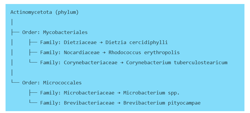
This means [35]:
Dietzia ↔ Rhodococcus = closest pair (same order, sister families)
Corynebacterium = moderately related (same order, different families)
Microbacterium & Brevibacterium = more distant (different order)
Mycobacteriales vs Micrococcales = major evolutionary separation
The first logical step is to remove the organisms in the order Micrococcales, based on fewer organisms within this order being recovered compared to the numbers recovered in the order Mycobacteriales ³⁵. Of the remaining organisms, it is logical to select either Dietzia or Rhodococcus based on their relative closeness and because they are recovered in far higher numbers than Corynebacterium (91% to 9%).
Gram-positive sporing rod selection
Gram-positive sporing rods are not often recovered. Given that B. subtilis is already included in the pharmacopoeia panel, the logical selection is B. cereus. Bacillus subtilis and Bacillus cereus belong to the same family (Bacillaceae), and both are Gram‑positive, spore‑forming bacilli, but they fall into two distinct evolutionary clades (monophyletic group) that diverged early in the genus Bacillus ³⁶. A clade is a grouping in biology that includes a common ancestor and all its descendants, representing a single, unbroken branch on the tree of life ³⁷:
Bacillus subtilis → “Subtilis clade”
Bacillus cereus → “Cereus clade”
These clades are separate, stable evolutionary lineages defined by conserved molecular signatures, as per Table 7 ³⁸.
Table 7: Clade associations of isolated Bacillus species
Feature | Bacillus subtilis | Bacillus cereus | Related? |
Family | Bacillaceae | Bacillaceae | ✔ Yes (broadly) |
Clade | Subtilis clade | Cereus clade | ✘ No (different clades) |
Evolutionary distance | Separate lineage with unique CSIs | Separate lineage with unique CSIs | ✘ Not close |
Sporulation | Yes | Yes | ✔ Shared ancient trait |
Ecological niche | Benign soil commensal | Soil; opportunistic human pathogen; foodborne | ❍ Different ecology |
Based on these differences, B. cereus is worthy of inclusion where a spore former is required.
Gram-negative rod selection
For Gram-negative rods, the following organisms appear across the different systems:
Pseudomonas fluorescens
Pseudomonas stutzeri
Moraxella osleonsis
Stenotrophomonas maltophilia
Moraxella plantain
Chryseobacterium indologenes
Of these organisms, the Moraxella bacteria were not found in high numbers. These are organisms linked to occasional poor personal hygiene (respiratory tract and face mask control). Of the other organisms, as noted above, Ps. fluorescens is a difficult culture to maintain due to its propensity to grow in cold storage. There are grounds to remove this bacterium based on practicality when preparing microbial cultures for dilution.
This leaves:
Pseudomonas stutzeri
Stenotrophomonas maltophilia
Chryseobacterium indologenes
These organisms are not closely related taxonomically ³⁹. The outcome is:
Chryseobacterium indologenes:
Belongs to phylum Bacteroidota.
Pseudomonas stutzeri and Stenotrophomonas maltophilia ⁴⁰:
Both belong to phylum Pseudomonadota (formerly Proteobacteria), specifically within a broader group of Gram‑negative non‑fermenters.
Historically, S. maltophilia was classified within Pseudomonas, then Xanthomonas, before being designated its own genus.
Stenotrophomonas is therefore more closely related to Pseudomonas than to Chryseobacterium.
All three organisms are:
Gram‑negative rods
Environmental bacteria, commonly found in water systems, soil, or moist surfaces.
Stenotrophomonas and Pseudomonas are common water-associated organisms, often recovered together in environmental monitoring.
Chryseobacterium spp. also appear frequently in environmental sampling, including water systems (e.g., C. indologenes, C. taichungense, C. soldanellicola).
Thus, even though they are phylogenetically distant, they co-occur in similar ecological niches, especially pharmaceutical water systems, cleanroom wet areas, and other low‑nutrient environments. Therefore, although there are differences, and based on each occurring at relatively similar numbers, any of the organisms is suitable for selection.
Fungi selection
Fungi are not commonly recovered. From the review, there are two species recovered in relatively higher numbers compared with other fungi:
Aspergillus fumigatus and Penicillium chrysogenum.
Both are filamentous fungi (both produce abundant airborne conidia, easily transferred by air and movement). It is noted that one filamentous fungus is included in the test panel - Aspergillus brasiliensis. However, the two Aspergilli are not closely related; they belong to different sections, represent distinct evolutionary lineages, and differ widely in ecology, pathogenicity, and physiology.
Aspergillus brasiliensis → member of the Nigri section (black Aspergilli), historically grouped with A. niger.
Aspergillus fumigatus → member of the Fumigati section, a thermotolerant species well-known for causing invasive aspergillosis.
These sections reflect deep phylogenetic separation within the genus (based on ITS, β‑tubulin, and calmodulin sequence comparison).
In considering the Penicillium, all three genera (Aspergillus and Penicillium) belong to the family Aspergillaceae, making them closely related at the family level, but they diverge into different genera and different evolutionary lineages ⁴¹,⁴². The outcome of this analysis is:
· Aspergillus brasiliensis → Section Nigri (black aspergilli)
· Aspergillus fumigatus → Section Fumigati (thermotolerant pathogenic group)
· Penicillium chrysogenum → Genus Penicillium, sister to Aspergillus within Aspergillaceae
And as illustrated in Figure 7.

This means:
A. brasiliensis and P. chrysogenum → moderately related
A. fumigatus and P. chrysogenum → moderately related (same degree)
A. brasiliensis vs A. fumigatus → more distantly related to each other than either is to Penicillium
It may seem counterintuitive, but A. brasiliensis and P. chrysogenum are closer to each other than either is to A. fumigatus. Hence, A. fumigatus is the appropriate fungus for selection.
Discussion
This paper has set out a method for considering the most appropriate environmental isolates to add to a test panel. This is based on a methodological approach that looks at numerical recoveries within different groupings and across different areas, followed by a vertical and horizontal genotypic comparison against the established pharmacopeial test panel and between the different isolates themselves.
Based on the above analysis, the recommended selection list for environmental isolates is as per Table 8.
Table 8: Final recommendations for additional organisms to include in the test panel
Type | Environmental isolate selection |
Gram-positive coccus | Either: Kocuria rhizophilia, Micrococcus luteus, K. carniphila, K. palustris |
Gram-positive non-sporing rod | Either: Dietzia cercidiphylli or Rhodococcus erythropolis |
Gram-positive sporing rod | Bacillus cereus |
Gram-negative rod | Either: Pseudomonas stutzeri , Stenotrophomonas maltophilia, Chryseobacterium indologenes |
Fungi | Aspergillus fumigatus |
Table 8 summarises the output from the review of multiple high‑signal systems (aseptic processing, process cleanrooms, water systems, and in‑process samples) to distil down a suitable index of organisms to complement the pharmacopeial test panel. This was achieved by selecting the most common isolates from a system first, then comparing for commonality between systems, and then ensuring potential isolates are sufficiently different from the compendial panel.
An alternative approach that could have been taken is to expand the criteria. Such an approach could consist of:
Frequency and prevalence (including the most recent trend).
Criticality of the source (where the organism was recovered from).
Patient or product impact (including the objectionable status of the organism).
Resistance profile of the organism to cleaning and disinfection agents.
Association of organisms with out-of-limits events.
Possible exclusion of organisms that are difficult to grow (with justification).
It is possible to weight and to score such factors and produce a total score to aid the final selection. A model ensures that isolates used for growth promotion testing, disinfectant efficacy studies, and method validation are representative of the facility microbiota, provide worst-case challenge conditions, and maintain appropriate morphological and ecological diversity.
Whichever approach is adopted, it needs to be practicable, scientifically justified, and regularly reviewed. The structured approach in this paper can aid microbiologists working in the pharmaceutical and healthcare sectors in developing their own approaches.
References
1. Salaman-Byron Facts about Environmental Isolates and Growth Promotion Test, American Pharmaceutical Review. May 30, 2019, at https://www.americanpharmaceuticalreview.com/Featured-Articles/359629-Facts-about-Environmental-Isolates-and-Growth-Promotion-Test/
2. Neidhardt, F. How microbial proteomics got started. Proteomics 2011; 11, 2943–2946
3. Dworkin, J., Shah, I. Exit from dormancy in microbial organisms. Nat Rev Microbiol 2010; 8, 890–896
4. Westney, R. The Role of In-House Microbial Isolates in Contamination Control. In Madsen, R. E. and Moldenhauer, J. (Eds.) Contamination Control in Healthcare Product Manufacturing, Volume 1, 2013. Parenteral Drug Association / DHI, U.S., pp. 475-504
5. Leak, R. E. and Leech, R. Challenge testing and their predictive ability. In Bloomfield, S. F., Baird, R., Leake, R. E. and Leech, R. (Eds.) Microbial Quality assurance in Pharmaceuticals, Cosmetics and Toiletries, 1988; Ellis Horwood, Chisterster, U.K., pp. 129-146
6. Sandle, T. Microbiological Culture Media: A Complete Guide for Pharmaceutical and Healthcare Manufacturers, 2018; DHI/PDA, Bethesda, MD, USA pp. 219-239
7. Booth, C. Environmental Isolates: What's The Proper Use Of In-House Cultures?, Pharmaceutical Online, 2019: https://www.pharmaceuticalonline.com/doc/environmental-isolates-what-s-the-proper-use-of-in-house-cultures-0001
8. Orth, D.S. Evaluation of preservatives in cosmetic products. In Kabara, J.J. (Ed.) Cosmetic and Drug Preservation: Principles and Practice, 1984; Marcel Dekker, New York, pp. 403-421
9. Grice, E., Kong, H., Renaud, G. et al. A diversity profile of the human skin microbiota. Genome Research. 2018; 18 (7): 1043–1050
10. Takarada, H., Sekine, M., Kosugi, H. et al. Complete genome sequence of the soil actinomycete Kocuria rhizophila. Journal of Bacteriology. 2008; 190 (12): 4139–46
11. Takeuchi M, Hatano K (Union of the genera Microbacterium Orla-Jensen and Aureobacterium Collins et al. in a redefined genus Microbacterium. Int J Syst Bacteriol. 1998; 48 (3): 739–747
12. Kanayama Y, Sakai Y. Purification and properties of a new type of protease produced by Microbacterium liquefaciens. Biosci Biotechnol Biochem. 2005;69(5):916-21
13. Sandle T The risk of Bacillus cereus to pharmaceutical manufacturing. American Pharmaceutical Review 2014; 17 (6): 56
14. Vilas-Bôas GT, Peruca AP, Arantes OM. Biology and taxonomy of Bacillus cereus, Bacillus anthracis, and Bacillus thuringiensis. Canadian Journal of Microbiology. 2007; 53 (6): 673–687
15. Garrido-Sanz, D.; Arrebola, E.; Martínez-Granero, F. et al. Classification of Isolates from the Pseudomonas fluorescens Complex into Phylogenomic Groups Based in Group-Specific Markers. Frontiers in Microbiology. 2017; 8: 413
16. Kloos WE, Musselwhite MS. Distribution and persistence of Staphylococcus and Micrococcus species and other aerobic bacteria on human skin. Appl Microbiol. 1975;30(3):381-5
17. Kaprelyants, AS, Kell, DB. Dormancy in stationary-phase cultures of Micrococcus luteus: flow cytometric analysis of starvation and resuscitation. Appl Environ Microbiol 1993; 59: 3187–3196
18. Kati, H., Ince, I.A., Demir, I., and Demirbag, Z. Brevibacterium pityocampae sp. nov., isolated from caterpillars of Thaumetopoea pityocampa (Lepidoptera, Thaumetopoeidae). Int. J. Syst. Evol. Microbiol. 2010; 60:312-316
19. Hinic V, Lang C, Weisser M. et al. Corynebacterium tuberculostearicum: a potentially misidentified and multiresistant Corynebacterium species isolated from clinical specimens. J Clin Microbiol. 2012;50(8):2561-7
20. Errington J, Aart LT Microbe Profile: Bacillus subtilis: model organism for cellular development, and industrial workhorse. Microbiology. 2020; 66 (5): 425–427
21. Alkhatib NJ, Younis MH, Alobaidi AS, Shaath NM. An unusual osteomyelitis caused by Moraxella osloensis: A case report. Int J Surg Case Rep. 2017;41:146-149
22. Andersen B, Frisvad JC, Søndergaard I. et al. Associations between fungal species and water-damaged building materials. Appl. Environ. Microbiol. 2011; 77 (12): 4180–8
23. Hördt A, López MG, Meier-Kolthoff JP. et al. Analysis of 1,000+ Type-Strain Genomes Substantially Improves Taxonomic Classification of 'Alphaproteobacteria'. Frontiers in Microbiology. 2020; 11: 468
24. Dworkin M, Falkow S, Rosenberg E. et al. The Prokaryotes, Volume 5 - Proteobacteria: Alpha and Beta Subclasses (3rd ed.). 2006; Springer. pp. 15–18
25. Kang YS, Kim J, Shin HD et al. Methylobacterium platani sp. nov., isolated from a leaf of the tree Platanus orientalis. Int J Syst Evol Microbiol. 2007;57(Pt 12):2849-2853
26. Rainey FA, Klatte S, Kroppenstedt RM, Stackebrandt E. Dietzia, a new genus including Dietzia maris comb. Nov., formerly Rhodococcus maris. Int J Syst Bacteriol. 1995; 45: 32–36
27. van der Geize R. and L. Dijkhuizen Harnessing the catabolic diversity of Rhodococci for environmental and biotechnological applications. Microbiology. 2004; 7 (3): 255–261
28. Bhuyar, G; Shah, H; Jain, S; Mehta, VK Urinary tract infection by Chryseobacterium indologenes. Indian Journal of Medical Microbiology. 2012; 30 (3): 370–2
29. Oren A, Garrity GM. Valid publication of the names of forty-two phyla of prokaryotes. Int J Syst Evol Microbiol. 2021; 71 (10): 5056
30. Williams KP, Kelly DP. Proposal for a new class within the phylum Proteobacteria, Acidithiobacillia classis nov., with the type order Acidithiobacillales, and emended description of the class
Gammaproteobacteria. International Journal of Systematic and Evolutionary Microbiology. 2013; 63 (Pt 8): 2901–2906
31. Varghese G, Jamwal A, Sarawat D. et al. Case Report: Case Series of Kocuria palustris Bacteremia among Immunocompromised Patients. Am J Trop Med Hyg. 2023 Sep 18;109(5):1113-1117
32. Kloos, W. E.; Schleifer, K. H. Isolation and Characterization of Staphylococci from Human Skin II. Descriptions of Four New Species: Staphylococcus warneri, Staphylococcus capitis, Staphylococcus hominis, and Staphylococcus simulans. International Journal of Systematic Bacteriology. 1975; 25 (1): 62–79
33. Nouioui I, Carro L, García-López M. et al. (2018). Genome-Based Taxonomic Classification of the Phylum Actinobacteria. Front. Microbiol. 2018; 9: 2007
34. Katı H, İnce İA, Demir İ, Demirbağ Z. Brevibacterium pityocampae sp. nov., isolated from caterpillars of Thaumetopoea pityocampa (Lepidoptera, Thaumetopoeidae). Int J Syst Evol Microbiol. 2010;60 (Pt 2):312-316
35. Mitchell BI, Markantonis JE. An underestimated pathogen: Corynebacterium species. J Clin Microbiol. 2025; 8;63(10):e0155224
36. Zhernakova DV, Wang D, Liu L et al. Host genetic regulation of human gut microbial structural variation, Nature. 2024;625(7996):813-821
37. Bhandari, V., Ahmod, N.Z., Shah, H.N. & Gupta, R.S. Molecular signatures for Bacillus species: demarcation of the Bacillus subtilis and Bacillus cereus clades in molecular terms and proposal to limit the placement of new species into the genus Bacillus. International Journal of Systematic and Evolutionary Microbiology, 2013;63(Pt 7):2712-2726
38. Pace, Norman R. Time for a change. Nature. 2006; 441 (7091): 289
39. Nikolaidis, M., Hesketh, A., Mossialos, D., et al. A Comparative Analysis of the Core Proteomes within and among the Bacillus subtilis and Bacillus cereus Evolutionary Groups Reveals the Patterns of Lineage- and Species-Specific Adaptations. Microorganisms, 2022; 10(9), 1720; https://doi.org/10.3390/microorganisms10091720
40. Majumdar R, Karthikeyan H, Senthilnathan V, Sugumar S. Review on Stenotrophomonas maltophilia: An Emerging Multidrug- resistant Opportunistic Pathogen. Recent Pat Biotechnol. 2022;16(4):329-354
41. Varga J, Kocsubé S, Tóth B et al. Aspergillus brasiliensis sp. nov., a biseriate black Aspergillus species with world-wide distribution. Int J Syst Evol Microbiol. 2007;57(Pt 8):1925-1932
42. Hong SB, Go SJ, Shin HD et al. Polyphasic taxonomy of Aspergillus fumigatus and related species. Mycologia. 2005;97(6):1316-29
Author
Corresponding Author: Tim Sandle, Head of Microbiology
Bio Products Laboratory,
UK Operations, England
Email: timsandle@btinternet.com


Comments